〒213-0002 神奈川県川崎市高津区二子1丁目7−17
リバーサイドマンション杉崎 102 二子新地駅 徒歩3分
| 月 | 火 | 水 | 木 | 金 | 土 | 日祝 | |
|---|---|---|---|---|---|---|---|
| 9:00〜13:00 | ● | ● | ● | ● | ● | ● | ─ |
| 15:00〜19:00 | ● | ● | ● | ● | ● | ● | ─ |
画像所見と症状の不一致について:腰椎椎間板ヘルニア
公開日:2026/01/09
更新日:2026/05/28
腰椎椎間板ヘルニアにおける画像診断の結果と実際の臨床症状が必ずしも一致しないという「解離」の実態を統計的に解説しています。
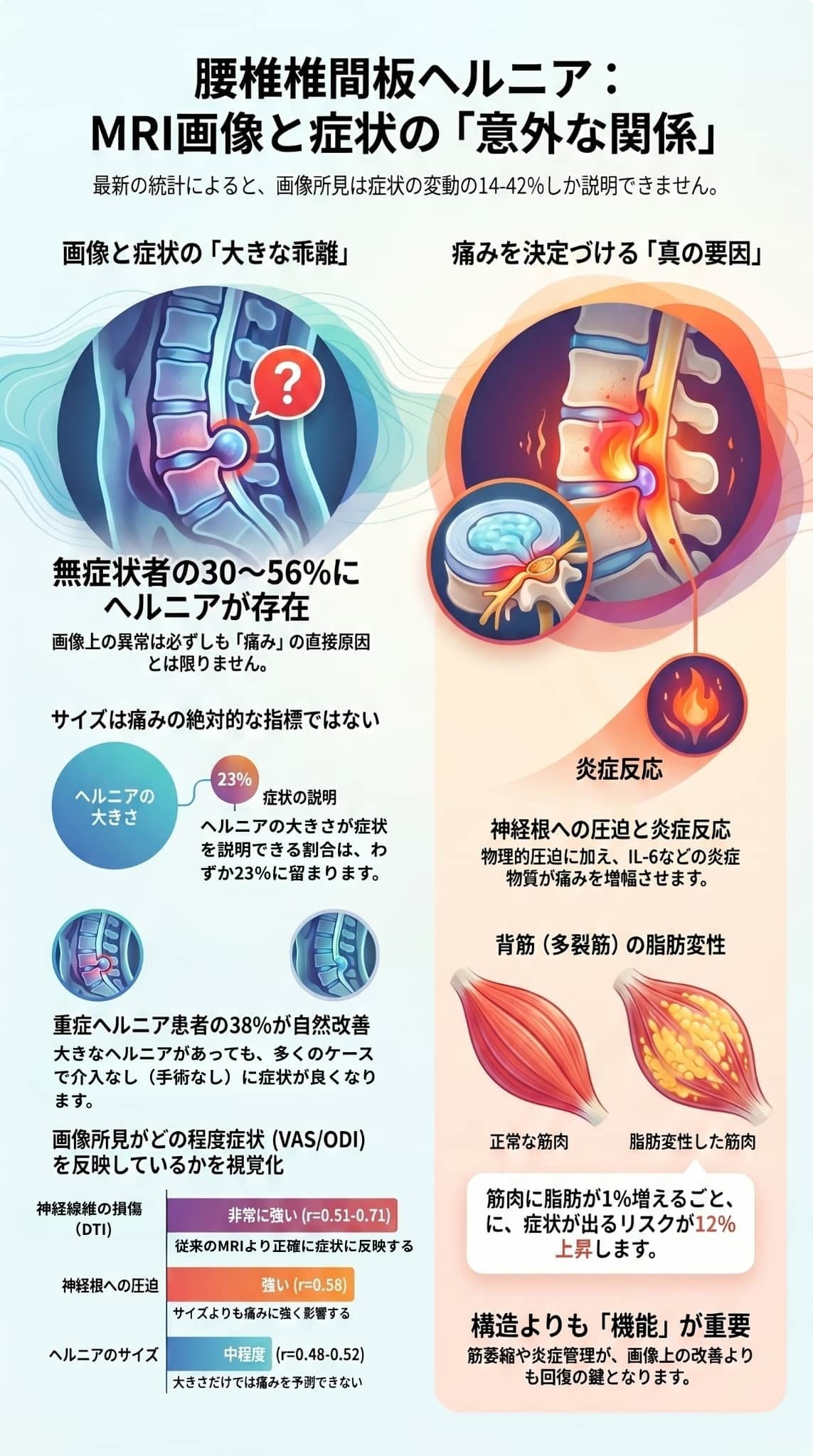
研究データによると、MRIで確認される異常が症状を説明できる割合は4割未満に過ぎず、健康な人の半数近くにもヘルニア所見が認められることが示されています。
症状の有無を左右するのは単なる突出の大きさではなく、神経根の圧迫度や炎症反応、さらには背筋の萎縮といった多角的な要因です。
そのため、画像のみに依存せず、保存療法やリハビリテーションを優先する包括的なアプローチが重要であると結論付けています。
最新の画像技術である**拡散テンソル画像(DTI)**を活用することで、より精密な神経損傷の評価が可能になる点も強調されています。
目次
研究の結果、MRIによる画像上の重症度と、実際の痛みや障害の程度との相関は、弱から中程度(r = 0.23-0.65)にとどまることが明らかになりました。
画像所見だけで症状の変動を説明できる割合はわずか14〜42%であり、残りの大部分は画像以外の要因(心理的要因や中枢性感作など)が関与していると考えられています。
症状のない健康な人であっても、30%から56%の割合で画像上に椎間板ヘルニアが認められるという事実が、複数の研究で示されています。これは、「画像に異常があるからといって、それが必ずしも症状の原因ではない」ということを示唆しており、画像診断の限界を浮き彫りにしています。
多変量解析の結果、単なるヘルニアの大きさよりも、以下の要素が症状の存在や重症度をより強く予測することが分かりました。
- 神経根の圧迫度: 面積の減少や変位の程度。
- 炎症バイオマーカー: 血清中のIL-6やTNFなどの炎症反応。
- 脊柱起立筋(多裂筋)の変性: 筋肉の萎縮や脂肪浸潤の割合。
- 終板の変化: Modic変化などの椎体終板の異常。
急性(12週未満)と慢性(12週以上)の症状では、画像および炎症のプロファイルが異なります。慢性患者は、より高度な椎間板変性、筋肉の萎縮、脂肪浸潤を示す傾向があり、これは単なるヘルニアの発生を超えた、二次的な適応変化が障害に関与していることを示しています。
レポートは、以下の結論を導き出しています。
- 画像所見は臨床的な文脈の中で解釈されるべきであり、画像のみに基づいて治療を決定してはなりません。
- ヘルニアの大きさだけで手術の必要性を判断するのは不十分です。
- 大きなヘルニアがあっても、約38%の患者は手術なしで自然に改善するため、原則として保存療法を第一に選択すべきです。
- 拡散テンソル画像(DTI)などの高度な画像技術は、従来のMRIよりも神経損傷の状態を正確に捉えられる可能性があります。
総じて、このレポートは腰椎椎間板ヘルニアにおける障害が多因子的であることを強調しており、画像、炎症、筋機能、そして心理社会的な側面を含めた包括的な評価の重要性を説いています。
腰椎椎間板ヘルニア(LDH)の有病率と人口統計的特性について、参考文献に基づいた詳細を以下にまとめます。
最も特筆すべき点は、症状のない健康な人々においても椎間板ヘルニアが高い割合で認められることです。
- 有病率の範囲: 無症状の人におけるヘルニアの有病率は、年齢や診断基準によって異なりますが、**30%から56%**に及びます。
- 加齢の影響: この割合は高齢になるほど高くなる傾向があります。
- 所見の内容: 無症状であっても、単なる突出(protrusion)だけでなく、より重度の脱出(extrusion)や神経根への接触が認められるケースもあります。
- 椎間板変性: ヘルニアに至らない「椎間板変性」のみであれば、無症状者における割合はさらに高くなります。
実際に症状がある人々の中では、その経過(急性か慢性か)によって画像上の特徴が異なります。
- 重症度の分布(手術症例): 手術を検討する患者群では、突出(protrusion)が28%、脱出(extrusion)が58%、遊離型(sequestration)が14%という分布が報告されており、より重度のタイプが過半数を占めています。
- 地域社会ベースの分布: 一方で、地域コミュニティベースの調査では、脱出よりも軽度な膨隆(bulge)や突出の割合が高くなります。
- 慢性化の影響: 慢性腰痛(12週以上)の患者は、急性患者と比較して高度な椎間板変性(グレード3以上)を示す割合が有意に高い(慢性:72% vs 急性:45%)ことが示されています。
- 年齢: 症状を伴うLDH患者の多くは30歳から60歳の間に集中しています。ある研究のコホートでは、平均年齢は52.3±11.7歳でした。
- 性別: 研究によってばらつきがあります。手術症例を対象とした研究では**男性が62%**と多い傾向にありましたが、地域ベースのサンプルでは男女比はより均衡しています。これは男性の方が職業的またはバイオメカニクス的なリスク因子にさらされやすいためと考えられています。
- BMI(肥満度): BMIが高いことは腰椎への機械的負荷を増加させますが、他の要因を調整した後の症状の重症度に対する独立した予測因子としては、統計的に一貫した結果が得られていません。
- 急性 vs 慢性: 急性症状(12週未満)と慢性症状(12週以上)では、筋肉の状態に明らかな差が見られます。慢性患者は、急性患者に比べて多裂筋の萎縮(断面積の減少)や脂肪浸潤が顕著です。
- 神経損傷の進行: 神経根の微細構造の変化を測定すると、症状の期間が長くなるほど神経損傷(FA値の低下)が進行するという有意な相関が認められています。
これらの統計データは、画像上の異常が必ずしも症状に直結しないこと(画像と症状の解離)を示しており、診断には臨床症状との慎重な照らし合わせが不可欠であることを裏付けています。
参考文献に基づき、腰椎椎間板ヘルニア(LDH)における有症状者(痛みやしびれがある人)と無症状者(健康な人)の比較について詳しく解説します。
主な違いは、単なるヘルニアの有無ではなく、**「筋肉の状態」「炎症反応」「神経の微細構造」**に現れています。
最も注目すべき点は、症状がなくても画像上はヘルニアが認められるケースが非常に多いことです。
- 無症状者の保有率: 健康で症状のない人の**30%〜56%**に椎間板ヘルニアが認められます。
- 年齢の影響: この割合は年齢とともに増加します。
- 所見の深刻度: 無症状の人でも、単なる「膨隆(bulge)」だけでなく、より重度の「脱出(extrusion)」や神経根への接触が見られることがあります。
- 結論: 画像上の異常は、症状を引き起こすための「必要条件」ではあっても「十分条件」ではないことが示唆されています。
有症状者と無症状者を分ける大きな要因の一つが、背骨を支える多裂筋(multifidus muscle)の状態です。
- 脂肪浸潤率: 有症状者は無症状者に比べて、筋肉内に脂肪が入り込む「脂肪浸潤」の割合が有意に高いことが示されています(有症状者:18.7% vs 無症状者:12.4%)。
- 筋肉の断面積(CSA): 有症状者(特に慢性患者)は、無症状者に比べて多裂筋の断面積が有意に減少(萎縮)しています。
- 予測因子: 多裂筋の脂肪浸潤率が1%上がるごとに、症状が出る確率が12%上昇するという分析結果もあります。
体内および局所の炎症状態も、両者の間で明確な差があります。
- 血清IL-6: 慢性的な症状がある患者は、無症状者に比べて血清中のインターロイキン-6(IL-6)レベルが有意に高くなっています(有症状者:8.4 pg/mL vs 無症状者:4.1 pg/mL)。
- 局所の炎症: 筋肉由来の炎症性サイトカイン(TNFなど)の発現も、有症状者でより顕著に認められます。
- 結論: 痛みなどの症状は、物理的な圧迫だけでなく、これらの化学的な炎症プロセスに強く依存していると考えられています。
Pfirrmann分類:椎間板変性の程度を示すます。
ヘルニアそのものよりも、椎間板全体の変性(老化・劣化)の度合いが症状の有無に関係しています。
- 変性グレード: 有症状者は無症状者に比べ、Pfirrmann分類による変性グレードが有意に高い傾向にあります。
- 慢性化との関連: 特にグレード3以上の高度な変性は、慢性的な腰痛を予測する独立した因子(オッズ比3.8)となっています。
最新の画像技術(DTI:拡散テンソル画像)を用いると、従来のMRIでは分からない神経の損傷状態の違いが見えてきます。
- FA値(異方性比): 神経が圧迫され症状が出ている部位では、無症状の部位や反対側に比べてFA値が有意に低くなっており、神経の微細構造に損傷が起きていることを示しています。
DTI(拡散テンソル画像法):MRI(磁気共鳴画像)技術の一つ。従来のMRIでは判別が難しい「神経根の微細なダメージ」や「生理的な圧迫の度合い」を定量的に評価できる最先端の手法。
FA値(分画異方性):MRIの拡散テンソル画像(DTI)で使われる指標
有症状者と無症状者を比較すると、**「ヘルニアの大きさ」よりも「周囲の筋肉の健康度」「炎症の有無」「神経の質的な損傷」**が、実際に症状が出るかどうかの境界線となっていることが統計的に明らかになっています。
参考文献に基づき、腰椎椎間板ヘルニア(LDH)における急性症状(12週未満)と慢性症状(12週以上)の比較について、画像所見、炎症、筋肉の状態、および神経損傷の観点から詳しく解説します。
主な違いは、単なるヘルニアの物理的な形状よりも、周囲の組織の二次的な変化や炎症のプロファイルに顕著に現れています。
急性期と慢性期では、MRIで観察される椎間板やヘルニアの質的特徴が異なります。
- 椎間板変性の程度: 慢性患者は、急性患者と比較して高度な椎間板変性(Pfirrmannグレード3以上)を示す割合が有意に高い(慢性:72% vs 急性:45%)ことが示されています。
- ヘルニアの質的特徴: 急性ヘルニアは慢性と比較して、T2信号強度が高く(水分の含有量や炎症を反映)、神経根の変位(圧迫による移動)が大きい(急性:4.2mm vs 慢性:2.8mm)傾向があります。
- 硬膜外造影増強: 急性症例では、慢性症例(18%)に比べて硬膜外の造影増強(42%)が多く見られ、活動的なプロセスが起きていることを示唆しています。
急性症状と慢性症状では、関与している炎症のメカニズムが異なります。
- 急性症状: 全身性の炎症マーカーである血清C反応性タンパク(CRP)が有意に高い(急性:12.3 mg/L vs 慢性:6.8 mg/L)ことから、より活動的な炎症反応が背景にあると考えられます。
- 慢性症状: 全身性よりも、局所(筋肉など)の炎症が目立ちます。慢性患者の多裂筋では、炎症性サイトカインであるTNFの発現が有意に上昇しており、筋肉由来の炎症が痛みの維持に関与している可能性があります。また、血清IL-6の高値も慢性の独立した予測因子となっています。
- 筋肉の萎縮: 慢性患者は、急性患者に比べて**多裂筋の断面積(CSA)が有意に減少(萎縮)**しています。
- 脂肪浸潤: 筋肉内に脂肪が入り込む「脂肪変性」も慢性期に顕著です。脂肪浸潤指数は、慢性患者(2.8)の方が急性患者(1.6)よりも有意に高いことが報告されています。
- 臨床的意義: これらの筋肉の変性は、単なるヘルニアの発生を超えた、慢性期特有の**「適応障害」**を反映していると考えられています。
- 微細構造の変化: 神経根の健康状態を示すFA値(異方性比)は、症状の期間が長くなるほど低下する(負の相関)ことが確認されています。
- 進行性損傷: これは、神経根への圧迫が長引くほど、神経の微細構造における損傷が進行することを示唆しています。
- 残存痛の予測: 手術前の症状持続期間が6ヶ月を超えていることは、手術後の残存痛(低バックペイン)の独立した予測因子となります。
- 治療アプローチ: 慢性患者では、画像上のヘルニアそのものへの対処だけでなく、筋肉の退行変性、中枢性感作、心理社会的要因などを考慮した、より包括的なアプローチが必要とされます。
| 比較項目 | 急性症状 (<12週) | 慢性症状 (≥12週) |
|---|---|---|
| 椎間板変性 | 比較的軽度 | 高度(グレード3以上)が多い |
| ヘルニアの特徴 | T2信号高値、神経変位大 | T2信号低下、神経変位小 |
| 主な炎症 | 全身性(CRP高値) | 局所筋肉(TNF高値)、血清IL-6高値 |
| 背筋(多裂筋) | 比較的健全 | 萎縮および脂肪浸潤が顕著 |
| 神経根の損傷 | 軽度から中等度 | 期間に比例して損傷が進行(FA値低下) |
このように、慢性期は**「単なる構造上の問題(ヘルニア)」から「筋肉や神経の質的変化、持続的な炎症」へと病態が移行している**のが大きな特徴です。
画像診断における腰椎椎間板ヘルニア(LDH)の重症度分類間の比較について、ソースに基づき、**「ヘルニアの形態(タイプ)」「椎間板の変性度」「神経根の圧迫度」**の3つの観点から解説します。
ヘルニアの形状や突出の程度に基づく分類(膨隆、突出、脱出、遊離)間の比較では、以下の傾向が示されています。
- 分布の差: 手術を要する患者群では、**脱出(extrusion)が58%**と最も多く、次いで突出(protrusion)が28%、遊離(sequestration)が14%を占めています。一方、地域社会ベースの調査では、突出や膨隆の割合がより高くなります。
- 痛みとの相関: 脱出や遊離は、突出に比べて平均痛みスコアが高い傾向にありますが、症状の程度には大きな個人差があり、各グループ間で重なり(オーバーラップ)が見られます。
- 診断の正確性: MRIによる形態分類は術中所見と強く相関(r = 0.78)しますが、術前の痛みスコアとの相関は中程度(r = 0.45)に留まります。
椎間板自体の劣化状態を示すPfirrmannグレード(1〜5)を用いた比較では、症状の慢性化との強い関連が認められます。
- 慢性化の指標: 慢性腰痛患者(12週以上)の72%がグレード3以上の高度変性を示したのに対し、急性患者では45%、無症状者では28%でした。
- 予測因子: 多変量解析において、**グレード3以上の変性は、慢性腰痛を予測する独立した因子(オッズ比 3.8)**であることが示されています。これは、単なるヘルニアの大きさよりも、椎間板全体の劣化が慢性的な障害に寄与していることを示唆しています。
ヘルニアの大きさそのものよりも、**神経根がどの程度圧迫されているか(断面積の減少率など)**の分類が、症状の重さをより強く反映します。
- 痛みスコア(VAS)の差: 神経根の圧迫を「軽度」「中等度」「高度(断面積が50%以上減少)」に分けた場合、高度圧迫群の平均VASスコアは7.8であり、中等度(6.2)や軽度(4.9)に比べて有意に高くなっていました。
- 機能障害(ODI)への影響: 多変量線形回帰分析において、神経根圧迫の重症度は、ヘルニアの大きさよりも機能障害(ODIスコア)を強く予測する因子(β = 8.4)として特定されています。
各分類を多変量モデルで比較すると、興味深い事実が明らかになっています。
- ヘルニアサイズ vs 圧迫度: 多くの研究において、単なる**「ヘルニアの大きさ」は、他の要因(神経圧迫度、筋肉の変性、炎症反応)を調整した後は、独立した症状予測因子として残らない**ことが報告されています。
- その他の重要因子: ヘルニアの重症度分類以外に、**「終板のModic変化」や「多裂筋の脂肪浸潤」**といった所見を組み合わせる方が、ヘルニアの形態分類単独よりも高い精度で症状や治療予後を予測できることが示されています。
画像診断上の重症度を比較すると、「ヘルニアの大きさや形態」よりも、「神経根の圧迫度」や「椎間板の変性グレード」の方が臨床症状をより強く反映することが統計的に裏付けられています。しかし、どの分類を用いても画像所見だけで症状のすべてを説明することはできず(説明率は14〜42%)、画像上の重症度は臨床的な文脈の中で解釈される必要があります。
参考文献に基づき、腰椎椎間板ヘルニア(LDH)における画像所見と臨床症状の全体的な相関関係について詳しく解説します。
結論から述べると、**画像上の重症度と実際の症状との相関は「弱から中程度」**であり、画像だけで症状のすべてを説明することは困難であることが統計的に示されています。
複数の研究を統合した分析の結果、画像所見(MRI)と症状(痛みや障害)の相関係数(r)は、一般的に 0.23 〜 0.65 の範囲に留まっています。
- 説明率(r²)の低さ: これは、画像所見が症状の変動を説明できる割合がわずか 14% 〜 42% であることを意味します。
- 未解明の要因: つまり、症状の 58% 〜 86% は画像以外の要因(心理的要因、中枢性感作、遺伝、社会的要因など)によって決まっていると考えられています。
画像と症状が必ずしも一致しないことを示す、象徴的な統計データがいくつかあります。
- 無症状者の高い保有率: 健康で痛みがない人でも、30% 〜 56% の割合で画像上に椎間板ヘルニアが認められます。
- 自然改善の存在: 大きなヘルニアがある患者のうち、38% は手術などの介入なしで症状が自然に改善します。
- 重症度の逆転: 逆に、画像上は軽微な異常しかなくても、激しい痛みや障害を抱える患者も存在します。
- 神経根の圧迫度: ヘルニアのサイズそのものよりも、神経根がどれだけ物理的に圧迫・変位しているかの方が、痛み(VAS)や障害(ODI)と強く相関します。
- 炎症バイオマーカー: 局所や血中の炎症反応(IL-6やTNFなど)の強さは、構造的な異常よりも痛みの強さを正確に反映する傾向があります。
- 多裂筋の変性: 背骨を支える筋肉(多裂筋)の萎縮や脂肪浸潤の度合いは、特に慢性症状の重症度と密接に関連しています。
- 神経根の微細な損傷を数値化するFA値は、症状の強さや持続期間とより強い相関(r = 0.51 〜 0.71)を示します,,。これは、物理的な形よりも「神経が受けている質的なダメージ」の方が症状に直結していることを示唆しています。
治療方針を決定する際には、画像所見のみに基づかず、患者の臨床症状や身体所見と照らし合わせて慎重に判断することが推奨されています。
参考文献に基づき、特定の画像所見がどのような臨床転帰(症状の持続、手術の必要性、術後の経過など)と関連しているかについて解説します。
統計的分析の結果、単なる「ヘルニアの大きさ」よりも、以下の特定の所見が患者の予後を予測する重要な因子であることが示されています。
以下の所見がある場合、保存療法で改善しにくく、手術が必要になる可能性や症状が持続するリスクが高まります。
- 後縦靭帯(PLL)の断裂: 手術を必要とした患者の62%に認められたのに対し、自然改善した患者では28%にとどまり、手術の必要性を予測する強い因子(オッズ比 4.1)となっています。
- Modic変化(椎体終板の信号異常): 症状の持続や手術の必要性と強く関連しており(オッズ比 3.4)、これがある場合は保存療法による改善が得られにくい傾向があります。
- 神経根の圧迫度: ヘルニアのサイズそのものよりも、神経根の断面積が50%以上減少するような「高度な圧迫」がある場合、痛み(VAS)や機能障害(ODI)が有意に重くなることが示されています。
手術(経皮的内視鏡下腰椎椎間板摘出術など)を受けた後、痛みがすっきり取れるかどうかに関わる因子です。
- 高輝度領域(HIZ): MRIで椎間板後方に白く写る小さな点は、術後の残存腰痛の独立した予測因子(オッズ比 2.9)となります。
- 術前のModic変化: これも術後の残存痛を予測する因子の一つです。
- 症状の持続期間: 術前に6ヶ月以上症状が続いていた場合、術後の回復が遅れたり痛みが残りやすくなったりします。
痛みが3ヶ月(12週)以上続く「慢性腰痛」への移行に関連する指標です。
- 多裂筋(背筋)の脂肪浸潤と萎縮: 筋肉内に脂肪が入り込み、筋肉の断面積が減少している場合、慢性的な痛みや障害の重症度と強く相関します。
- 高度な椎間板変性(Pfirrmannグレード3以上): 慢性患者の72%に認められ、急性患者(45%)よりも有意に高い割合です。
- 自然改善の割合: 画像上大きなヘルニアがあっても、患者の約38%は手術なしで臨床的に大幅な改善を示します。
- 画像と症状の不一致: ヘルニアの形態分類(突出や脱出など)と術前の痛みスコアの相関は中程度(r = 0.45)であり、画像がひどく見えても予後が良い、あるいはその逆というケースが頻繁に起こります。
- FA値(異方性比): 拡散テンソル画像(DTI)で測定される神経根のFA値は、従来のMRIよりも症状の重症度と強く相関(r = -0.71)します。
- 回復の指標: 6ヶ月後のフォローアップにおいて、**FA値の改善は臨床的な症状改善と強く相関(r = 0.64)**しており、神経の微細構造の回復が良好な転帰の指標となります。
臨床転帰を判断する上では、ヘルニアの「形」だけでなく、**「靭帯の損傷」「終板の変化(Modic変化)」「筋肉の状態」「神経の微細なダメージ(FA値)」**を総合的に評価することが、より正確な予測につながると結論づけられています。
参考文献に基づき、腰椎椎間板ヘルニア(LDH)における画像所見と臨床症状の**「不一致(解離)のパターン」**について詳しく解説します。
近年の研究では、画像上の異常と実際の症状が一致しないケースが頻繁に認められており、主に以下の4つのパターンに分類されます。
画像と症状の関係は一方向ではなく、以下の両方のパターンが存在します。
- 画像は重度だが無症状(あるいは軽症): 大きなヘルニアがあっても、臨床的に大幅な改善を示す患者が約38%存在します。
- 画像は軽微だが重症: 初期のMRIでは比較的小さなヘルニアしか認められないにもかかわらず、症状が持続または悪化する患者が約12%存在します。
最も顕著な不一致のパターンは、全く症状のない健康な人にヘルニアが認められるケースです。
- 高い保有率: 無症状者の30%〜56%に椎間板ヘルニアが認められ、その割合は加齢とともに上昇します。
- 所見の種類: 単なる膨隆(bulge)だけでなく、脱出(extrusion)や神経根への接触といった重度の所見が無症状者に現れることもあります。
- 結論: 画像上の異常は症状が出るための「必要条件」かもしれませんが、それだけで症状を引き起こす「十分条件」ではないことを示しています。
- 画像上の鮮度と症状: 一般に急性ヘルニアはT2信号が高く、神経の変位も大きいという特徴がありますが、こうした「急性の画像特徴」を持ちながら「慢性の症状」を抱えている患者、あるいはその逆のケースも確認されています。
- 自然経過: 症状が完全に消失した後も、画像上はヘルニアが依然として残っているという時間的なズレもしばしば認められます。
- 14〜42%の説明力: 従来の画像診断(ヘルニアの大きさや型)が症状の変動を説明できる割合は、統計的にわずか14%〜42%に過ぎません。
- 残りの58〜86%: 症状の大部分(約6割〜9割)は、画像には写らない以下の要因によって決まっていると考えられています。
- 心理的要因: 破局的思考、恐怖回避思考など。
- 中枢性感作: 痛みの伝達系が過敏になる現象。
- 炎症プロファイル: 物理的圧迫以外の化学的な刺激。
- 社会的要因: 職場環境や生活背景。
参考文献に基づき、腰椎椎間板ヘルニア(LDH)における症状の有無や重症度を予測する独立因子について詳しく解説します。
多裂筋の健康状態は、症状の発生と慢性化を予測する極めて重要な因子です。
- 脂肪浸潤率: 多裂筋への脂肪浸潤率が1%上昇するごとに、有症状となるリスクが12%上昇する(オッズ比 1.12)ことが示されています。
- 筋肉の断面積(CSA): 多裂筋の断面積が大きいことは、症状の発生に対して**保護的な役割(予防的効果)**を果たします。逆に、筋肉の萎縮(CSAの減少)は慢性腰痛の独立した予測因子となります。
ヘルニアの形状そのものよりも、神経が受けている物理的・質的なダメージが症状に直結します。
- 神経根の圧迫度: 神経根の断面積の減少率や変位の程度は、痛み(VAS)や障害(ODI)の重症度を予測する強力な因子です。
- 神経の微細構造の変化(DTI指標): 拡散テンソル画像(DTI)で測定されるFA値(異方性比)の低下は、従来のMRI指標よりも症状の強さや期間を正確に予測します。
全身的および局所的な炎症反応の強さが、痛みの存在を予測します。
- 血清IL-6レベル: 血中のインターロイキン-6(IL-6)濃度の上昇は、慢性腰痛の独立した予測因子です(1 pg/mL上昇につきオッズ比 1.15)。
- 局所の炎症: 筋肉由来のTNFなどのサイトカイン発現も、構造的な異常以上に症状の強さと相関します。
- Modic変化(椎体終板の信号異常): 症状が持続し、手術が必要になるリスクを大幅に高める因子(オッズ比 3.4)です,。また、術後の残存痛の予測因子でもあります。
- 後縦靭帯(PLL)の断裂: 靭帯の断裂が認められる場合、保存療法で改善せず手術が必要になる確率が4倍以上高まります(オッズ比 4.1)。
- 椎間板変性グレード: Pfirrmannグレード3以上の高度な変性は、慢性腰痛を予測する独立した因子(オッズ比 3.8)となります。
- 高輝度領域(HIZ): MRIで椎間板後方に認められる高輝度領域は、術後の残存痛を予測する因子となります。
- 症状の期間: 術前に6ヶ月以上症状が持続していることは、不良な臨床転帰の予測因子となります。
特筆すべき統計的結論として、「ヘルニアの大きさや形態」は、上記の因子(筋肉、炎症、神経圧迫、終板変化)を調整した後は、症状の有無を予測する独立した因子として残らないケースが多いことが報告されています。
つまり、治療方針や予後の判断においては、単なる「ヘルニアが出ているか」だけでなく、**「筋肉が衰えていないか」「神経が物理的にどれほど圧迫されているか」「周囲に炎症や終板の変性があるか」**を総合的に評価することが重要であると結論づけられています。
参考文献に基づく多変量回帰分析の結果、腰椎椎間板ヘルニア(LDH)における症状の存在や重症度を予測する**「独立因子(他の要因の影響を除いても関連が認められる因子)」**について解説します。
統計学的な分析において、多くの人が重視しがちな**「ヘルニアの大きさや形態」は、他の要因を考慮した後は独立した予測因子として残らない**ケースが多いことが明らかになっています。
代わりに、以下の5つの要素が症状の重症度をより正確に予測する因子として特定されています。
ヘルニアのサイズそのものよりも、神経が受けている物理的・質的なダメージが直接的な予測因子となります。
- 圧迫の深刻度: 神経根の断面積が50%以上減少するような高度な圧迫は、痛み(VASスコア)や機能障害(ODIスコア)の重症度を予測する強力な因子です,,。多変量モデルにおいて、神経根の圧迫度はヘルニアのサイズよりも高い予測値(β = 8.4)を示します。
- DTI指標(FA値): 拡散テンソル画像(DTI)で測定されるFA値(異方性比)の低下は、従来のMRI指標よりも症状の強さ(r = -0.71)や持続期間を正確に予測する独立した指標となります。
背骨を支える多裂筋の状態は、症状の発生と慢性化の主要な決定因子です。
- 脂肪浸潤率: 多変量ロジスティック回帰分析では、多裂筋の脂肪浸潤率が1%上昇するごとに、有症状となるリスクが12%上昇する(オッズ比 1.12)ことが示されています。
- 筋肉の断面積(CSA): 多裂筋の断面積(筋肉の太さ)は症状に対して保護的に働き、逆に**筋肉の萎縮(CSAの減少)**は慢性腰痛の独立した予測因子となります。
- 血清IL-6レベル: 血中のインターロイキン-6(IL-6)の上昇は、慢性腰痛を予測する独立した因子です(1 pg/mLの上昇につきオッズ比 1.15)。
- 局所のTNF発現: 筋肉由来のTNF(腫瘍壊死因子)などのサイトカイン発現は、構造的な異常以上に痛みのスコアと強く相関(r = 0.64)します。
- Modic変化: 椎体終板の信号異常(Modic変化)は、症状の持続(オッズ比 3.4)や手術後の残存痛(オッズ比 3.6)を予測する強力な因子です。
- 後縦靭帯(PLL)の断裂: 靭帯の断裂が認められる場合、保存療法で改善せず手術が必要になるリスクが4倍以上高まります(オッズ比 4.1)。
- 高輝度領域(HIZ): MRIで椎間板後方に認められる小さな高輝度スポットも、術後の残存痛の独立した予測因子(オッズ比 2.9)です。
- 6ヶ月以上の持続: 手術前に症状が6ヶ月以上続いていることは、治療後の回復が遅れたり、痛みが残りやすくなったりすることを示す独立した予測因子となります。
参考文献に基づき、腰椎椎間板ヘルニア(LDH)における症状の有無や重症度を決定する要因の比較結果について解説します。
統計学的な多変量解析を総合すると、単なる「ヘルニアの大きさ」よりも、神経根の圧迫度、筋肉の状態、炎症反応、および終板の変化の方が、症状を説明する上で遥かに重要な決定要因であることが示されています。
主要な決定要因の比較分析結果は以下の通りです。
- 圧迫の重要性: 複数の研究において、神経根の圧迫度(断面積の減少、変位、信号変化など)は、ヘルニアのサイズ単独よりも強力な予測因子として一貫して示されています。
- 統計的有意性: 多変量モデルにおいて、他の要因(圧迫度など)を調整した後では、ヘルニアの大きさそのものは統計的な有意性を失うことが頻繁にあります,。これは、大きなヘルニアがあっても神経根をうまく避けていれば症状が出にくいことを示唆しています。
- 独立した決定要因: 多裂筋の萎縮(断面積の減少)や脂肪浸潤は、症状の存在と重症度の両方における独立した決定要因です。
- バイオメカニクス的意義: 筋肉の健康状態は、脊柱の安定性というバイオメカニクス的要因と、神経損傷に伴う二次的な変化(神経原性変化)の両方を反映しており、臨床転帰を左右する極めて重要な要素と見なされています。
- 炎症の影響力: 全身性(血清CRP、IL-6など)および局所(筋肉内のTNFなど)の炎症マーカーは、症状と独立して関連しています。
- 痛みの本質: 炎症マーカーの方が構造的な画像所見よりも症状と強く相関するケースがあり、LDHの痛みは単なる「機械的な圧迫」よりも「化学的な炎症プロセス」に密接に関連していると考えられています。
- 予後の予測因子: 椎体終板のModic変化や高輝度領域(HIZ)は、症状の持続性や治療(手術)後の転帰を予測する上で、椎間板ヘルニアそのものよりも臨床的に関連性が高い場合があります。
- 持続的な痛みの原因: これらは進行中の炎症や変性プロセスを反映しており、痛みが長引くかどうかの重要な指標となります。
- 神経へのダメージ: 症状の持続期間は、神経根の微細構造的なダメージ(DTIで測定されるFA値の低下)と負の相関があります。
- 慢性化のメカニズム: 期間が長くなるほど筋肉の変性が進み、適応障害が生じるため、単なるヘルニアの形態以上に回復の難しさを決定づける要因となります。
比較分析の総括として、ヘルニアの大きさや形態は、基礎となる病態の「マーカー(目印)」に過ぎない可能性があり、実際の臨床症状を直接決定づけているのは、神経根への実質的なダメージ、局所の炎症、そして周囲の筋機能の低下であると結論づけられています。
参考文献に基づき、2016年から2026年にかけての腰椎椎間板ヘルニア(LDH)に関する**統計的証拠の統合(Synthesis of Statistical Evidence)**について、主要な5つの結論を解説します。
最も重要な証拠の一つは、症状のない健康な人であっても30%から56%の割合で椎間板ヘルニアが認められるという事実です。
- この高い有病率は、画像上の異常があるだけで診断や治療計画を立てることの妥当性に根本的な疑問を投げかけています。
- MRIで見られるヘルニアは、患者の臨床症状と解剖学的・時間的に一致する場合にのみ臨床的意義を持つと解釈すべきです。
統計分析により、画像上の重症度と実際の症状(痛みや障害)の相関は、一貫して「弱から中程度(r = 0.23〜0.65)」であることが示されています。
- これは、画像所見が症状の変動を説明できる割合がわずか**14%〜42%**に過ぎないことを意味します。
- 残りの大部分(58%〜86%)は、心理的要因、中枢性感作、遺伝、社会的要因など、画像には写らない多因子的要素によって決まっています。
- 神経根の圧迫度: 物理的な変位や断面積の減少。
- 脊柱起立筋(多裂筋)の変性: 筋肉の萎縮や脂肪浸潤の程度。
- 炎症バイオマーカー: 血清中のIL-6レベルや局所のサイトカイン。
- 終板の変化: Modic変化や高輝度領域(HIZ)。
- 慢性患者: 高度な椎間板変性、顕著な筋肉の萎縮、脂肪浸潤を伴う傾向があります。
- 急性患者: 全身性の炎症反応(CRP高値)や、画像上での高いT2信号強度(水分の多さ)が特徴的です。
- この違いは、慢性化が初期のヘルニア発生を超えた、二次的な適応障害を含んでいることを示唆しています。
- DTIで測定されるFA値(異方性比)などは、神経根の「形」ではなく「微細構造的なダメージ」を反映しており、これが症状の強さや持続期間をより正確に捉えています。
参考文献に基づき、腰椎椎間板ヘルニア(LDH)における統計的分析結果が示唆する**臨床的意義(診断や治療にどのように活かすべきか)**について解説します。
- 症状との照らし合わせ: 無症状者の30〜56%にヘルニアが認められるため、MRIで見える異常は、それが患者の症状と解剖学的および時間的に一致する場合にのみ臨床的に意味を持ちます。
- 偶発的所見の除外: 症状のない部位で見つかったヘルニア(偶発的所見)に基づいて治療を決定すべきではありません。
- 神経根の状態を優先: ヘルニアの形よりも、**神経根の圧迫度や微細な損傷状態(DTIによる評価など)**の方が、症状の強さや予後をより正確に反映します。
- 周囲組織のチェック: 脊柱起立筋(多裂筋)の萎縮や脂肪浸潤、および椎体終板のModic変化は、症状の持続や手術の必要性を予測する強力な因子です,。これらをルーチンの評価に組み込むべきです。
- 自然改善の可能性: 大きなヘルニアがあっても、患者の約38%は特別な介入なし(手術なし)で自然に改善することが示されています。
- 手術の適応: 手術は、進行性の神経学的欠損、馬尾症候群、または適切な保存療法を行っても改善しない深刻な障害がある場合に限定すべきです。
- 二次的変化への対処: 慢性期では、単なるヘルニアの物理的な問題だけでなく、筋肉の変性、中枢性感作(痛みに敏感になること)、心理社会的な要因が複雑に絡み合っています。
- リハビリの重要性: 筋肉の健康状態が症状を予測することから、理学療法や運動療法、神経筋肉のリハビリテーションは治療の重要な構成要素となります。
- 破局的思考の防止: 画像上の異常を強調しすぎると、患者が過度に不安を感じ(破局的思考)、恐怖から活動を避ける行動(恐怖回避思考)を招き、結果として障害を長引かせる可能性があります。
- 正しい理解の共有: 「健康な人でも画像に異常が出ることは珍しくない」という事実を伝え、画像上の異常が必ずしも将来の痛みや重症度を決定するものではないことを理解してもらうことが重要です。
臨床医は「画像(MRI)を治療する」のではなく、画像・臨床症状・炎症状態・筋機能、そして心理的側面を統合した「患者全体」を評価して治療方針を立てるべきである、というのが最新の統計的証拠が示す結論です,。
2016年から2026年にかけての統計研究を統合した本レポートの結論は、以下の5つの主要なポイントに集約されます。
腰椎椎間板ヘルニア(LDH)における画像上の重症度と臨床症状の相関は、統計的に**「弱から中程度(r = 0.23-0.65)」にとどまります,,,。従来のMRIによる画像所見だけで症状の変動を説明できるのは全体の14〜42%**に過ぎず、残りの大部分は心理的要因、中枢性感作、遺伝的背景などの非画像要因によって決まっています。
多変量解析の結果、単なるヘルニアの大きさや形態よりも、以下の要素が症状の有無や重症度を予測する上でより強力な独立した決定要因であることが証明されました。
- 神経根の圧迫度: 物理的な圧迫による断面積の減少や変位の程度。
- 多裂筋(背筋)の変性: 筋肉の萎縮や脂肪浸潤の割合。
- 炎症バイオマーカー: 血清中のIL-6レベルや、局所のTNFなどの炎症反応。
- 終板の変化: Modic変化などの椎体終板の異常。
- 症状中心の解釈: 画像はあくまで補助診断とし、身体所見や症状と解剖学的に一致する場合にのみ臨床的意義を認めます。
- 保存療法の優先: 進行性の神経損傷がない限り、自然改善の可能性を考慮して保存療法を第一選択とすべきです。
- 多面的なケア: 慢性期には、画像上の構造異常だけでなく、筋機能の回復(リハビリ)、炎症の制御、心理的教育を組み合わせた包括的なアプローチが不可欠です。
参考文献にに基づくと、拡散テンソル画像(DTI)T2マッピングといった最新の画像技術は、従来のMRIでは捉えきれなかった**「神経根の微細な構造変化」や「組織損傷のバイオマーカー」を数値化できる**という点で非常に優れています。
- FA値(異方性比)の低下: 神経が圧迫され、損傷が起きている部位では、健全な部位に比べてFA値が有意に低くなることが示されています。
- ADC値(見かけの拡散係数)の上昇: 反対に、神経の損傷に伴ってADC値は上昇する傾向があります。
- DTIの指標(特にFA値)は、機能障害の指標であるODIスコアや痛みの強さと**強い負の相関(r = -0.51 〜 -0.71)**を示します。
- これは、従来のMRI指標による相関(r = 0.38 〜 0.65)よりも高く、より**臨床的に関連性の高い「神経損傷のバイオマーカー」**として機能することを示唆しています。
- 損傷の進行: 症状の期間が長くなるほどFA値が低下する傾向があり、長期間の圧迫によって神経の微細構造的なダメージが進行している様子が分かります。
- 回復の予測(予後予測): 治療開始から6ヶ月後の追跡調査において、FA値の改善と臨床的な症状の改善が強く相関している(r = 0.64)ことから、治療がうまくいっているかの客観的な指標(予後予測因子)としての価値も認められています。
- T2マッピング: DTIに加えて、T2マッピングなどの高度なMRIシーケンスは、組織損傷の新たなバイオマーカーを提供し、診断の精度を高めるために活用されています。
[1] Chen, Y., et al., "Evidence of MRI image features and inflammatory biomarkers association with low back pain in patients with lumbar disc herniation," The Spine Journal, 2024, doi: 10.1016/j.spinee.2024.02.006.
[2] Wang, X., et al., "The effect of lumbar multifidus muscle degeneration on upper lumbar disc herniation," Frontiers in Surgery, 2024, doi: 10.3389/fsurg.2024.1323939.
[3] Dutta, S., et al., "Correlation of 1.5 Tesla Magnetic Resonance Imaging with Clinical and Intraoperative Findings for Lumbar Disc Herniation," Asian Spine Journal, vol. 10, no. 6, pp. 1115-1122, 2016, doi: 10.4184/ASJ.2016.10.6.1115.
[4] Šprláková-Puková, A., et al., "MRI Predictors for Improvement Without Any Intervention of Clinical Symptoms in Patients With Lumbar Disc Herniation, Questioning the True Need for Surgery," Radiology Research and Practice, 2026, doi: 10.1155/rrp/4954622.
[5] Hu, Z., et al., "Predictive factors for residual low back pain following percutaneous endoscopic lumbar discectomy in patients with lumbar disc herniation," Medical Science Monitor, 2023, doi: 10.12659/msm.942231.
[6] Hasanovic-Vučković, L., et al., "Correlation Between MRI Findings, ODI, and VAS Score in Lumbar Disc Herniation," Sarajevo Medical Journal, 2024, doi: 10.70119/0021-24.
[7] mou, X., et al., "Correlation between MRI manifestations of lumbar disc herniation and fatty infiltration of paravertebral muscles and chronic low back pain," Proceedings of the International Society for Magnetic Resonance in Medicine, 2024, doi: 10.58530/2024/1551.
[8] Aizawa, T., et al., "New diagnostic support tool for patients with leg symptoms caused by lumbar spinal stenosis and lumbar intervertebral disc herniation: a self-administered, self-reported history questionnaire," Journal of Orthopaedic Science, 2016.
[9] Sima, A., et al., "Reconsidering high intensity zones: its role in intervertebral disk degeneration and low back pain," European Spine Journal, 2024, doi: 10.1007/s00586-024-08185-x.
[10] Erbüyün, K., et al., "Lumbar Disc Herniation, the Association Between Quantitative Sensorial Test and Magnetic Resonance Imaging Findings," Pain Medicine, vol. 19, no. 11, pp. 2235-2244, 2018, doi: 10.1093/PM/PNX236.
[11] Saini, V., et al., "Clinical Reporting of Magnetic Resonance Imaging, the Way Forward for Patients With Lumbar Disc Herniation: A Prospective Correlational Study," Cureus, 2022, doi: 10.7759/cureus.27232.
[12] Divi, S. N., et al., "Can Imaging Characteristics on Magnetic Resonance Imaging Predict the Acuity of a Lumbar Disc Herniation," The International Journal of Spine Surgery, 2021, doi: 10.14444/8032.
[13] Erbüyün, K., et al., "Lumbar Disc Herniation, the Association Between Quantitative Sensorial Test and Magnetic Resonance Imaging Findings," Pain Medicine, vol. 19, no. 11, pp. 2235-2244, 2018, doi: 10.1093/pm/pnx236.
[14] Rijal, L., et al., "Correlation between Clinical Features and Findings Observed on Magnetic Resonance Imaging in Patients with Lumbar Disc Prolapse," 2020.
[15] Chen, Y., et al., "Applicative value of T2 mapping in evaluating lumbosacral nerve root injury induced by lumbosacral disc herniation," Acta Radiologica, 2022, doi: 10.1177/02841851221128377.
[16] Raudner, M., et al., "Prediction of Lumbar Disk Herniation and Clinical Outcome Using Quantitative Magnetic Resonance Imaging: A 5-Year Follow-Up Study," Investigative Radiology, 2019, doi: 10.1097/RLI.0000000000000527.
[17] Abdallah, A., et al., "Factors associated with the recurrence of lumbar disk herniation: non-biomechanical–radiological and intraoperative factors," Neurological Research, 2022, doi: 10.1080/01616412.2022.2116525.
[18] Rahman, M. M., et al., "Association between clinically diagnosed lumbar intervertebral disc prolapse and magnetic resonance image findings," Bangabandhu Sheikh Mujib Medical University Journal, vol. 9, no. 3, pp. 140-145, 2016, doi: 10.3329/BSMMUJ.V9I3.29459.
[19] Yu, P., et al., "Prognostic factors for residual symptoms following percutaneous endoscopic lumbar discectomy," Neurosurgical Review, 2024, doi: 10.1007/s10143-024-02486-x.
[20] Lê, V. T., et al., "Tương quan của hình ảnh cộng hưởng từ với triệu chứng lâm sàng của bệnh thoát vị đĩa đệm cột sống thắt lưng," Tạp chí Điện quang Việt Nam, vol. 41, pp. 195-201, 2020, doi: 10.55046/vjrnm.41.195.2020.
[21] Pesonen, J., et al., "Extending the straight leg raise test for improved clinical evaluation of sciatica: validity and diagnostic performance with reference to the magnetic resonance imaging," BMC Musculoskeletal Disorders, 2021, doi: 10.1186/S12891-021-04649-Z.
[22] Eguchi, Y., et al., "Diffusion tensor imaging of radiculopathy in patients with lumbar disc herniation: preliminary results," The Bone & Joint Journal, vol. 98-B, no. 3, pp. 387-394, 2016, doi: 10.1302/0301-620X.98B3.36036.
[23] Fonseka, R. D., et al., "What Degree of Radiological Compression Predicts Cauda Equina Syndrome: A Retrospective Study at a National Tertiary Center," 2021.
[24] Eguchi, Y., et al., "Diffusion tensor imaging of radiculopathy in patients with lumbar disc herniation: preliminary results," Journal of Bone and Joint Surgery-British Volume, vol. 98-B, no. 3, pp. 387-394, 2016, doi: 10.1302/0301-620X.98B3.36036.
[25] Waldrop, R., et al., "The Association Between Magnetic Resonance Imaging Disc Pathology and Provocative Discography at the Lumbar Level," Journal of Computer Assisted Tomography, 2021, doi: 10.1097/RCT.0000000000000956.
[26] Phan, T. H., et al., "Prognosis of disc herniated patients using back pain functional scale," Vietnamese Journal of Neurology, 2024, doi: 10.62511/vjn.42.2024.027.
[27] El-Hady, S. A., et al., "Evaluation of health related quality of life with the use of Oswestry disability index in degenerative discogenic low back pain," Egyptian Rheumatology and Rehabilitation, 2023, doi: 10.1186/s43166-022-00166-6.
[28] Singh, R., et al., "A Prospective Study to Evaluate the Clinical and Diffusion Tensor Imaging (DTI) Correlation in Patients with Lumbar Disc Herniation with Radiculopathy," Spine Surgery and Related Research, 2022, doi: 10.22603/ssrr.2022-0159.
[29] Antohe, I., et al., "The Relationship between Magnetic Resonance Imaging and Functional Tests Assessment in Patients with Lumbar Disk Hernia," Healthcare, vol. 11, no. 19, 2023, doi: 10.3390/healthcare11192669.
[30] Wu, W., et al., "Microstructural Changes in Compressed Nerve Roots Are Consistent With Clinical Symptoms and Symptom Duration in Patients With Lumbar Disc Herniation," Spine, vol. 41, no. 11, pp. E661-E666, 2016, doi: 10.1097/BRS.0000000000001354.
ごあいさつ

長引いた痛みを一人で治すのは困難なことが多いです。
困ったときは自身で判断せずに適切な処置を受けるために専門家に相談しましょう。
もし、お近くにお住まいで、困っているならば、一度ひまわり接骨院までお問い合わせください。腰痛・坐骨神経痛の専門家の新幡が、ご相談に乗ります。
気軽にご相談ください。

新着情報・お知らせ
お気軽にお問合せください

お電話でのお問合せ・相談予約
<受付時間>
月~土
9:00〜13:00 /15:00〜19:00
※日曜・祝日は除く
フォームは24時間受付中です。お気軽にご連絡ください。
ひまわり接骨院
住所
〒213-0002
神奈川県川崎市高津区二子1丁目7−17 リバーサイドマンション杉崎 102
アクセス
二子新地駅 徒歩3分
駐車場:近隣にコインパーキングあり。自転車・バイクは店舗前に駐輪場がございます。
受付時間
月~土
9:00〜13:00 /15:00〜19:00
定休日
日曜・祝日