〒213-0002 神奈川県川崎市高津区二子1丁目7−17
リバーサイドマンション杉崎 102 二子新地駅 徒歩3分
| 月 | 火 | 水 | 木 | 金 | 土 | 日祝 | |
|---|---|---|---|---|---|---|---|
| 9:00〜13:00 | ● | ● | ● | ● | ● | ● | ─ |
| 15:00〜19:00 | ● | ● | ● | ● | ● | ● | ─ |
頚椎症の重症度を決めるのは?
公開日:2026/07/02
更新日:2026/00/00
加齢に伴い首の神経が圧迫される頚椎症について、最新の研究成果に基づき患者向けに解説した包括的なガイドです。
病気の重症度や手術後の回復を予測するための重要な指標として、MRI画像に見られる脊髄の信号変化や、症状が続いている期間、さらには糖尿病などの持続的な持続疾患の影響を詳しく説明しています。
特に、早期の発見と適切なタイミングでの治療介入が、機能回復の鍵を握ることが強調されています。
また、人工知能を用いた最新の診断技術や、心の健康状態が予後に与える影響についても触れ、医療チームと協力して最適な治療法を選択するための具体的な助言を提示しています。
最終的に、患者が自らの状態を深く理解し、主体的にリハビリテーションや生活習慣の改善に取り組むことの重要性を説く内容となっています。
頚椎症(変性性頚椎脊髄症)の重症度および治療・手術後の経過(予後)を予測する主要な因子は、画像所見、臨床的特徴、そしてそれらを組み合わせた統合評価に大別されます。
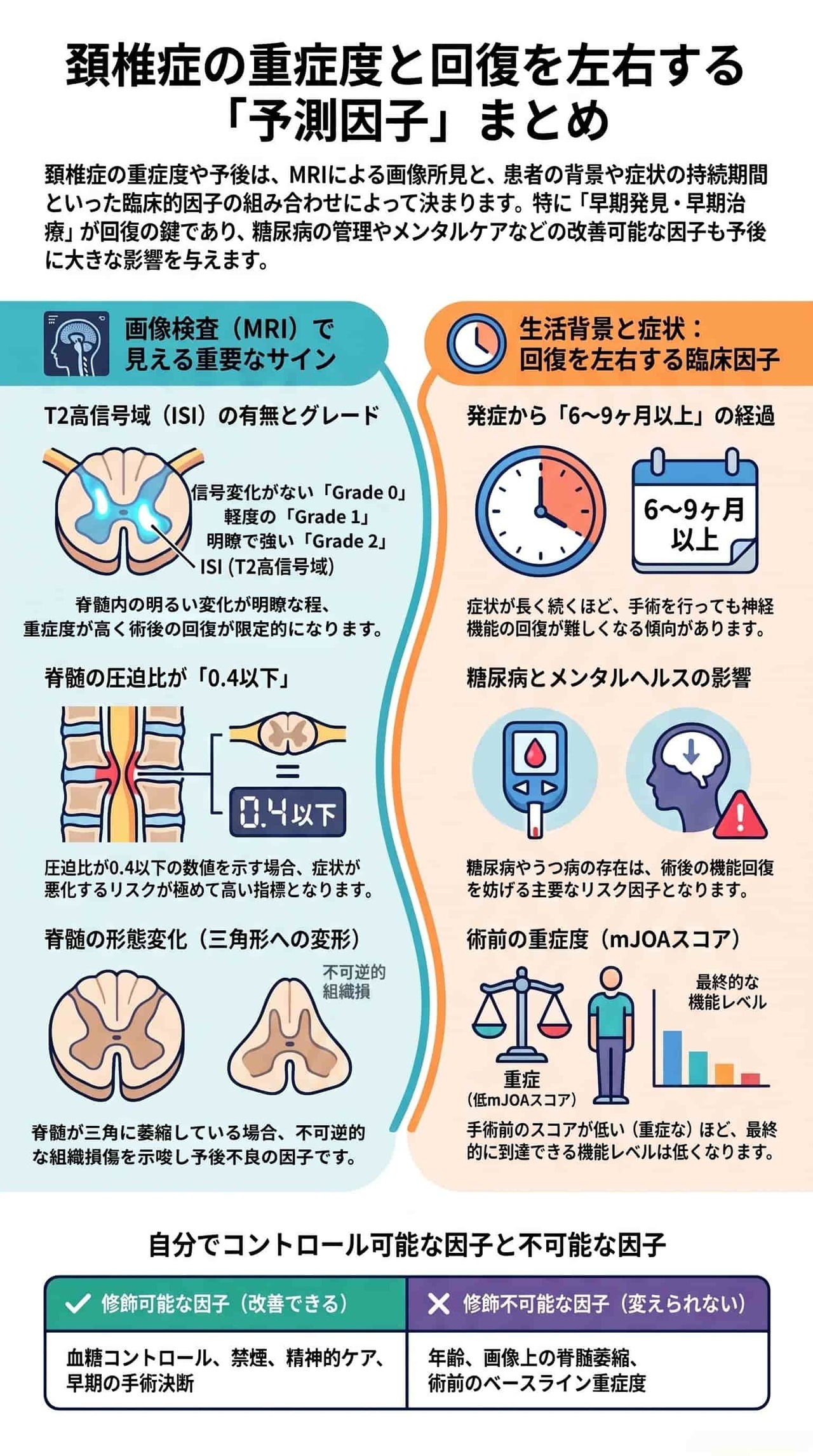
- T2高信号域(ISI)の有無とグレード: MRIのT2強調画像において、脊髄内部が明るく映る変化を指します。明瞭で強い信号変化(Grade 2)や、複数椎体にわたる広範囲の信号変化、あるいは信号強度比が1.455以上である場合、より重度な脊髄症や手術後の不良な経過(不良転帰のリスクが4.24倍に増加)を強力に予測する指標となります。
- 脊髄の圧迫度と横断面積: 圧迫部位における脊髄の前後径を左右径で割った**「圧迫比」が0.4以下**、または脊髄横断面積(CSA)が70.1 mm²以下の場合、無症状であっても将来的に症状を伴う脊髄症へ進行するリスクが高いとされています。また、前後両方向からの圧迫(双方向性圧迫)があるレベルの数が多いことも回復を鈍らせる要因です。
- 脊髄萎縮: 長期にわたる圧迫によって脊髄が細くなり、MRI上で三角形の形状を呈している場合、不可逆的な組織損傷(脊髄萎縮)が疑われ、術後の回復が限定的になる可能性を示唆します。
- 先進的な画像指標(DTIとDBSI): 研究段階の技術ですが、神経線維の整合性を反映するDTI(拡散テンソル画像)のFA値が0.5未満の患者は回復率が低い傾向(0.5以上が57.32%に対し、0.5未満は38.78%)にあります。さらに、軸索の微細な拡散状態を測る**DBSI(拡散基底スペクトル画像)と臨床指標を組み合わせたモデルでは、手術後の回復を90.5%**という高精度で予測できることが報告されています。
- 症状の持続期間(最も重要な修飾可能な因子): 発症から治療(手術)までの期間が長いほど、脊髄が不可逆的なダメージを受けるため予後が不良になります。具体的には6.5ヶ月以上(あるいは9ヶ月以上)続いている場合に予後不良リスクが高まり、1ヶ月長引くごとに不良な術後予後のオッズが約1.2倍ずつ増加します。早期に医療介入し、この待機期間を短縮することが非常に重要です。
- 初診時の重症度(mJOAスコア): 手指の動作、歩行、感覚、膀胱機能を数値化するmJOAスコア(18点満点)において、術前の重症度が高い(スコアが低い)患者ほど、術後改善の絶対値(点数の伸び幅)は大きくなる傾向にあります。しかし、最終的に到達できる機能レベル自体は、術前に軽症だった患者の方が良好です。
- 年齢: 高齢患者(特に70歳以上)は、若年患者に比べて術後の機能改善(mJOAスコアの改善度)が小さい傾向にあります。しかし、高齢であっても手術によって症状の進行を食い止め、生活の質(QOL)を改善させる効果は十分に実証されているため、高齢だけを理由に治療を諦めるべきではありません。
- 併存疾患(糖尿病・心血管疾患): 糖尿病は術後回復を妨げる顕著なリスク因子であり、多数の研究(52論文中85%)で不良な手術転帰との関連が指摘されています。また、心血管疾患も術後の神経学的回復を損なう傾向にあります。これらは、術前にコントロールを最適化することで悪影響を軽減できる可能性があります。
- 精神的健康(うつ病と不安): 術前の中等度〜重度のうつ病リスクは、術後の障害や痛みの改善を著しく阻害し、不良な経過をたどるリスクを1.36〜2.26倍(自己申告によるうつ病では約3倍)に高めることが報告されています。
単一の要因だけでなく、初期のmJOAスコア、性別、症状の持続期間、併存疾患などの臨床データを組み合わせることで、**約74.3%〜75%**の精度で手術後の機能悪化や良好な転帰を予測するシステムを構築できます 。
**頚椎症(変性性頚椎脊髄症:DCM)**は、首の骨や椎間板の加齢に伴う変化によって、脊髄や神経根が圧迫されることで引き起こされる病気です。成人における後天性脊髄障害の最も一般的な原因であり、特に55歳以上の方に多く見られます。
- 頚椎症性脊髄症(CSM): 首の骨の中を通る太い神経の束である「脊髄」が圧迫される状態です。手のしびれや歩行困難などの症状が現れ、進行性の経過をたどることが多いのが特徴です。
- 頚椎症性神経根症(CSR): 脊髄から枝分かれして手や腕に向かう「神経根」と呼ばれる細い神経が圧迫される状態です。
主な原因は加齢による首の構造変化です。骨の間でクッションの役割をしている椎間板が変性したり、骨にトゲのような突起(骨棘)が形成されたり、骨をつなぐ靭帯が厚くなったりすることで、神経の通り道が狭くなり圧迫が生じます。
この神経への圧迫は、常に圧迫されている「静的要因」と、首を動かしたときに一時的に生じる「動的要因」の両方によって引き起こされます。
また、首を動かすたびに神経に加わる微小な損傷や、血流低下による虚血(血流不足)も、神経障害を引き起こす原因となります。
- 手の細かい動作(巧緻動作)の障害: ボタンがうまくかけられない、文字が書きにくいなど。
- 歩行のふらつき: 歩くときに足元が不安定になり、つまずきやすくなるなど。
- 感覚異常・しびれ: 手足のしびれや、触った感覚が鈍くなる状態。
- 首の痛み
- 排尿障害: 重症化した際に見られることがあります。
自然経過の研究では、経過観察を行っている患者の57%が将来的に神経学的な悪化を示すことが報告されています。そのため、手のしびれや歩行のふらつきといったサインに早く気づいて専門医を受診し、適切なタイミングで治療(手術や保存療法)を行うことが、長期的な障害を防ぐために極めて重要です。
画像検査(主にMRI)によって、脊髄の圧迫の程度、内部のダメージ状態、および脊髄の形状変化といった構造的・機能的特徴を詳細に把握し、重症度や手術後の回復可能性を予測することができます。
- 3段階のグレード分類: 信号変化がない「Grade 0」、軽度な「Grade 1」、明瞭で強い変化がある「Grade 2」に分類されます。グレードが高いほど脊髄症は重症であり、手術による回復率も低くなります。
- 範囲と境界: 複数の骨のレベルにわたる広範囲の信号変化や、明瞭な境界を持つ信号変化は、その後の経過が良くない(予後不良)サインとされます。
- 信号強度比: この比率が1.455以上である場合、手術後の回復が不良となり、不良転帰(経過が良くないこと)のリスクが4.24倍に増加することが報告されています。
- 圧迫比: 圧迫されている部分の脊髄の前後径を左右径で割った値です。この値が0.4以下に低下していると、現在は症状がなくても将来的に手足のしびれなどの症状を伴う脊髄症へ進行するリスクが高くなります。
- 脊髄横断面積(CSA): 脊髄の断面の広さを表し、これが70.1 mm²以下に減少していることも脊髄症を発症する独立した予測因子になります。
- 双方向性圧迫のレベル数: 前後の両方向から脊髄が挟まれるように圧迫されている箇所の数が多いほど、手術後の改善が鈍くなります。
- 長期間にわたって強い圧迫を受け続けると、脊髄が細くなる「脊髄萎縮」が起こります。
- MRI画像上で脊髄が三角形の形状を呈している場合、これは萎縮が進んだ不可逆的な変化(神経の消失)を意味し、手術を行っても十分な機能回復が得られにくい傾向を示します(ただし、この予測的価値については研究間で一部見解が異なる部分もあります)。
- DTI(拡散テンソル画像)のFA値: 神経線維のつながり具合を評価する指標です。この値が低いほど損傷が進んでいることを示し、0.5未満の患者では術後の平均回復率が38.78%(0.5以上では57.32%)と低くなることが示されています。
- DBSI(拡散基底スペクトル画像): 神経線維とその周囲のダメージを区別して測定でき、手術後の改善予測を高い精度で行えます。臨床データと組み合わせることで、手術後の改善を**90.5%**という極めて高い精度で予測できることが報告されています。
**症状や患者背景(臨床的因子)**は、頚椎症の重症度や手術後の回復(予後)を予測する上で、画像検査と同じかそれ以上に重要な指標となります。これらは患者さん自身で把握できたり、治療前にコントロール(修飾)できたりする要素が多いのが特徴です。
症状が始まってから手術などの治療を受けるまでの期間は、その後の回復に最も強く影響します。
- 「6.5ヶ月」および「9ヶ月」の壁: 症状の持続期間が6.5ヶ月以上になると予後不良(回復が鈍くなる)の可能性が高まり、手術までの期間が1ヶ月長引くごとに、予後不良のオッズが約1.2倍ずつ増加します。また、持続期間が9ヶ月以上になると、不良な転帰をたどるリスクが約4倍に跳ね上がることが報告されています。
- 長期にわたり脊髄が圧迫され続けると不可逆的な(元に戻らない)損傷につながるため、手のしびれや歩行のふらつきなどのサインに気づいたら、早期に受診・治療して「待機期間」を短縮することが極めて重要です。
治療を始める前の神経障害の程度は、術後の最終的な到達点を予測する重要な基準となります。評価には、手指の動作や歩行、感覚、膀胱機能を数値化する「mJOAスコア(18点満点)」が用いられます。
- 改善の幅と最終的な機能のギャップ: 術前のスコアが低い(重症である)患者さんほど、手術によって改善する「絶対値(点数の伸び幅)」自体は大きくなる傾向があります。しかし、最終的に到達できる機能レベル(生活の便利さなど)は、術前に軽症(スコアが高かった)だった患者さんの方が良好になります。このため、重症化する前の早期介入が推奨されます。
年齢は手術後の機能改善の程度に影響を与えます。
- 若年者と高齢者の比較: 70歳以上の高齢患者と70歳未満の若年患者を比較した研究では、どちらのグループも手術後に有意な機能改善を示したものの、改善幅は若年群(mJOAスコアで平均3.8点改善)の方が高齢群(平均2.6点改善)よりも大きいことが示されています。高齢になるほど、関与する頚椎のレベル(骨の箇所の数)や症状の数が多くなり、術前の重症度が高くなりやすいことも影響しています。
- ただし、高齢であっても手術によって病気の進行を食い止め、生活の質(QOL)を改善する効果は十分に実証されているため、高齢であることを理由に治療を諦めるべきではありません。
- 糖尿病: 糖尿病に関する52の論文のうち85%(44論文)が不良な手術転帰との関連を報告しており、特に下肢の感覚や運動機能の回復を妨げる傾向があります。術前の徹底した血糖コントロールが推奨されます。
- 心血管疾患: 心血管疾患のある患者さんは、手術前後の合併症(周術期罹患率)のリスクが高く、神経学的な回復も不良になりやすいことが指摘されています。
- 喫煙: 喫煙は血流を低下させ組織の治癒を妨げるため、術前の重症度を高める要因や、手術後の「首の痛み」の改善を妨げるリスク因子になります。速やかな禁煙が推奨されます。
- うつ病の影響: 中等度〜重度のうつ病リスクがある場合、術後の障害や痛みの経過が不良になるリスクが1.36〜2.26倍に増加します。また、「自分はうつ病である」と自覚している患者さんでは、術後の障害が残るリスクが約3倍に増加することが報告されています。
- うつや不安は痛みの感覚を強め、リハビリへの意欲を低下させてしまいますが、これらは治療(精神療法や薬物療法など)によって改善可能な因子です。治療前にこうした心のケアを行うことが、全体の予後改善につながります。
頚椎症において「手術を受けるべきか、それとも保存療法で様子を見るべきか」という治療方針を決定する際は、まず**「脊髄症(CSM)」か「神経根症(CSR)」かという病態(タイプ)の違いと、それぞれの経過を左右する解剖学的・臨床的な予後予測因子**を総合的に評価します。
脊髄(首の骨の中を通る神経の束)が圧迫されているのか、それとも神経根(枝分かれして手や腕に向かう神経)が圧迫されているのかによって、放置した場合の自然経過が大きく異なるため、これが最も基礎となる決定因子です。
- 頚椎症性脊髄症(CSM)の場合:手術を優先的に検討
- 悪化のリスク: 脊髄症の自然経過は一般的に不良です。経過観察を行っている新規診断患者の57%(過去に手術を受け再発した患者では73%)が、平均2.5年の間に神経学的な悪化を示します。
- 治療基準: 中等度から重度の症例では、症状の進行を食い止め、生活の質(QOL)や機能を改善するために手術が強く推奨されます。軽症(mJOAスコア 15-17点)であっても、進行性の神経学的徴候や症状が見られる場合は手術介入が推奨されます。
- 頚椎症性神経根症(CSR)の場合:保存療法を第一選択として検討
- 自然経過の良さ: 神経根症の自然経過は比較的良好であり、強化運動やストレッチなどの理学療法(保存療法)によって約80%の患者が12週間以内に改善を示します。
- 手術を検討する基準: 保存療法を4〜6週間行っても症状が持続する場合や、進行性の悪化(特に手や腕の明らかな筋力低下など)がある場合は手術が有効です。特に椎間板ヘルニアを伴う患者では、手術によって首や腕の痛みが顕著に改善されることが報告されています。
- 自然経過の良さ: 神経根症の自然経過は比較的良好であり、強化運動やストレッチなどの理学療法(保存療法)によって約80%の患者が12週間以内に改善を示します。
- 神経孔(神経の出口)の狭窄が軽度: 画像検査において、神経孔の狭窄が軽度(最小斜位椎間孔面積が陰性レベル)な患者さんは、非手術治療によって首の痛みや手の障害、JOAスコアが大幅に改善することが示されています。逆に狭窄が顕著(陽性レベル)な場合は、保存療法での改善が限定的になります。
- 頚椎のアライメント(骨の並び方)が良好: 頚椎の前弯が十分に保たれている状態(大きなC2-7 Cobb角)の患者さんは、首の良好なバランスによって神経根への圧迫が自然と軽減されやすく、保存療法での良好な改善と関連していることが報告されています。
- 症状の持続期間が短い(12ヶ月未満): 首や腕の痛みが始まってから12ヶ月未満の段階で手術を受けた患者さんは、保存療法を選択した患者さんよりも、腕の痛みにおいて有意に良好な回復を示します。
- ※なお、脊髄症(CSM)においても、症状持続期間が6.5ヶ月〜9ヶ月以上になると術後回復が限定的になるため、これ以上のダメージを防ぐために早期手術(待機時間の短縮)が極めて重要です。
- ※なお、脊髄症(CSM)においても、症状持続期間が6.5ヶ月〜9ヶ月以上になると術後回復が限定的になるため、これ以上のダメージを防ぐために早期手術(待機時間の短縮)が極めて重要です。
- 健康関連QOL(生活の質)が低い: 日常生活の質を示す指数(EQ-5D)が著しく低下している患者さんでは、手術治療が首の痛みに対して非常に有効です。
- 精神的健康状態(不安やストレス): 首や腕の痛みに対する不安レベルが高い患者、日常生活のストレスレベルが高い患者、また「自分で痛みをコントロールできる」という感覚(自己効力感)が低い患者では、手術治療が痛みや障害の改善に対して特に高い効果を示します。
- 女性: 神経根症患者において、女性は手術を受けることで特に良好な経過(腕の痛みの改善)を示すことが確認されています。
保存療法で様子を見ている最中であっても、以下の症状や変化が現れた場合は、神経の不可逆的(元に戻らない)な損傷を防ぐために、速やかに専門医を受診し手術を検討すべきです。
- 手の握力の低下(症状悪化の早期検出に最も有用)
- 手指の巧緻動作の低下(ボタンがかけにくい、文字が書きにくいなど)
- 歩行のふらつきやバランス感覚の悪化(特につまずきやすさの増加や不整地での歩行困難)
- 排尿障害の出現(脊髄圧迫の重症化を示すサイン)
これらの予後予測因子を医療チームと共有し、ご自身の現在の状態(病態のタイプ、画像上の特徴、症状の持続期間など)と照らし合わせることで、より納得のいく治療方針を決定する(共同意思決定を行う)ことができます。
頚椎症性脊髄症(CSM)において、手術を受けるべきか保存療法で経過を見るべきかを決定し、さらに術後の回復(予後)の可能性を予測するための主要な因子は以下の通りです。
- 高い自然悪化率: 新規に診断されたCSM患者の57%(過去に手術を受け、その後に再発した患者では73%)が、平均2.5年の経過観察中に神経学的に悪化します。このため、保存療法の自然経過は一般的に不良であることを念頭に置く必要があります。
- 重症度に応じた推奨: 中等度から重度の症例(mJOAスコア12点未満など)では手術が強く推奨されます。軽症(mJOAスコア 15-17点)であっても、経過観察中に進行性の神経学的徴候や症状が見られる場合は手術介入が推奨されます。
保存療法を選択して様子を見ている最中であっても、以下の臨床的な変化が現れた場合は、手術へと切り替える強力な判断基準(予測因子)となります。
- 悪化の早期検出指標: 経過観察中の悪化を捉える際、mJOAスコア単独(感度33%)や通常の解剖学的MRI(感度28%)だけでは検出感度が低く、悪化を見逃すリスクがあります。代わりに**「握力の低下」「手の巧緻性(細かい動作)の低下」「歩行安定性の悪化」**を定量的にモニタリングすることが、悪化のサインを捉える早期検出指標として極めて有用です。
手術を選択した後に、実際にどの程度機能が回復するかを予測する指標です。これらを総合的に評価することで、手術に踏み切るべきかの期待値を算出できます。
- 症状の持続期間(最も重要な修飾可能因子): 症状が始まってから手術を受けるまでの期間は、術後予後を左右する最も重要な要素です。持続期間が6.5ヶ月以上で予後不良の可能性が高まり(1ヶ月追加されるごとに不良な予後のオッズが約1.2倍増加)、9ヶ月以上になると不良な転帰をたどるリスクが約4倍に跳ね上がります。脊髄の不可逆的な損傷を防ぐため、早期の手術介入(待機時間の短縮)が強く求められます。
- 術前の重症度(mJOAスコア): 術前のスコアが低い(重症な)患者ほど、手術による機能改善の絶対値(点数の伸び幅)は大きくなる傾向にあります。しかし、最終的に到達できる機能レベル自体は、術前に軽症だった患者の方が良好になります。
- 画像検査(MRI所見): MRIのT2強調画像での**T2高信号(特にGrade 2の明瞭で強い信号変化)**は、脊髄内のむくみや血流不足、不可逆的な組織損傷を反映し、手術後の不良な成績を予測する陰性因子です。また、強い脊髄圧迫比(0.4以下など)、頚椎後弯、多椎体レベルでの双方向性圧迫、脊髄内病変の長さなども手術後の回復を妨げる因子です。
- 併存疾患の管理状況: 糖尿病、高血圧、喫煙、抗凝固薬・抗血小板薬の使用は術後の不良な経過と関連します。特に糖尿病は下肢の回復を妨げる傾向がありますが、術前に適切にコントロール(最適化)することで影響を軽減できます。
- 年齢: 70歳以上の高齢患者であっても手術によって症状の進行を食い止め、生活の質(QOL)を改善させることが実証されていますが、70歳未満の若年群(mJOAスコアで平均3.8点改善)に比べると、高齢群(平均2.6点改善)は改善幅が小さくなる傾向があります。
頚椎症性神経根症(CSR)は、頚椎症性脊髄症(CSM)に比べて自然経過が比較的良好であり、強化運動、ストレッチ、牽引療法などの保存療法によって約80%の患者が12週間以内に改善します。 そのため、原則としてまずは保存療法が第一選択となります。
画像検査や首の構造的な特徴から、以下の条件を満たしている場合は、保存療法で十分に改善する可能性が高いため、手術を急がず様子を見る良い候補となります。
- 神経孔(神経の出口)の狭窄が軽度(画像所見): MRI等において「最小斜位椎間孔面積」の狭窄が顕著ではない(陰性)患者は、保存療法により手の障害(NDI)が平均10.32減少し、JOAスコアが平均2.86増加し、首の痛み(VAS)が平均2.46点改善するなど、大幅な回復が期待できます。逆に狭窄が顕著(陽性)な場合は、保存療法による改善は極めて限定的(NDIの減少が2.35、JOA増加が0.88にとどまる)になります。
- 頚椎のアライメント(骨の並び方)が良好: 頚椎の前弯が十分に保たれている状態(大きなC2-7 Cobb角)の患者は、首の良好なバランスによって神経根への圧迫が自然と軽減されやすく、保存療法による良好な改善と関連していることが報告されています。
- 症状の持続期間が短い(12ヶ月未満): 首や腕の痛みが始まってから12ヶ月未満の段階で手術を受けた患者は、保存療法を選択した患者よりも、特に腕の痛みにおいて有意に良好な回復を示します。
- 女性: 女性患者は、手術を受けることで腕の痛みの改善において特に良好な経過を示すことが確認されています。
- 健康関連QOL(生活の質)が著しく低い: 日常生活の質を示す指数(EQ-5D)が非常に低い患者では、手術治療が首の痛みに対して極めて高い効果を発揮します。
- 不安やストレスレベルが高い: 首や腕の痛みに対する不安レベルが高い、あるいは日常生活のストレスレベルが高い患者は、手術を行うことで首の痛みや障害が特に大きく改善します。
- 自己効力感(セルフエフィカシー)が低い: 「自分で痛みをコントロールできる」という感覚が低い患者は、手術を受けることで障害の程度が有効に改善します。
※この研究において、理学療法単独の方が手術よりも良好な転帰と関連する因子は見つかりませんでした。
様子を見る(保存療法)か手術を受けるかを決定するにあたり、以下のタイミングに関する因子も重要です。
- 手の脱力感や筋力低下(運動麻痺)の長期化: 手や腕の筋力低下(脱力)が長く続いている場合、手術を行っても運動機能の回復が困難になる傾向があります。そのため、保存療法を4〜6週間行っても症状が持続する場合や、進行性に悪化する場合は、MRI検査を含む専門的評価を受ける必要があります。
- 手術待機時間による悪化リスク: 手術を受けることが決まった場合、待機時間が長引くほど術後(2年後)の障害や痛みのすべてにおいて経過が不良になります。具体的には、手術が90日遅れるごとに、不良な転帰をたどるリスクが1.31〜1.64倍に増加します。
- 精神的健康状態(うつ病): 中等度〜重度のうつ病リスクがある場合、術後の回復が阻害され、不良な経過をたどるリスクが1.36〜2.26倍(自己申告によるうつ病がある場合は約3倍)に増加します。これらは治療(心理療法や薬物療法など)によって術前に改善可能な因子です。
頚椎症の経過管理や治療の成否には、患者さんとご家族による主体的な関与が極めて重要な役割を果たします。 科学的根拠に基づき、自宅での自己管理から医療チームとの連携まで、実践できる具体的なアドバイスを5つの柱に分けて紹介します。
頚椎症の症状はゆっくり進行するため初期に見過ごされやすく、診断までに平均して2.2〜6.3年も遅れることがあります。以下の症状が現れた場合、または保存療法(様子見)を4〜6週間続けても改善せず悪化する場合は、速やかに専門医(整形外科や脳神経外科)を受診し、MRI検査などの精密評価を受ける必要があります。
- 手の警告サイン: 手指のしびれ、異常な感覚、手の脱力感や握力の低下。
- 注: 運動麻痺(筋力低下)が長期化すると、手術を行っても回復しにくくなります。
- 注: 運動麻痺(筋力低下)が長期化すると、手術を行っても回復しにくくなります。
- 足の警告サイン: 歩行時のふらつき、階段の上り下りが困難になる、よくつまずく。
- 注: 単なる足の筋力低下と思い込み、腰の問題と決めつけずに首の検査を受けることが重要です。
- 注: 単なる足の筋力低下と思い込み、腰の問題と決めつけずに首の検査を受けることが重要です。
- その他の重症サイン: まれですが、排尿障害(おしっこが出にくい、コントロールできない等)は脊髄圧迫が極めて深刻になっているサインです。
保存療法で経過を観察している患者さんの**57%**が、平均2.5年の間に神経学的に悪化することが報告されています。医師の診察(mJOAスコア評価など)やMRI検査だけでは、日常の微妙な悪化を十分に捉えきれない(感度が約30%と低い)ため、自宅での継続的なセルフチェックが重要です。
- 握力の変化を測る: 握力は脊髄の悪化を早期に見つける極めて実用的な指標です。
- 手指の「巧緻性(細かい動作)」をチェックする: 「ボタンがかけづらい」「文字が書きにくい」「お箸が使いづらい」といった変化がないか確認します。
- 歩行の安定性を測る: 10秒間ステッピングテストや30メートル歩行テストなど、自宅でもできる簡単な物理テストで足の動きを客観的に記録します。また、暗い場所やでこぼこした道で歩きにくくなっていないか注意を払います。
- アドバイス: これらの変化をご家族が優しく観察・記録し、受診時に具体的な日常生活の変化として医師に伝えることで、適切な手術タイミングの決定に大きく役立ちます。
予後(治療後の経過)を悪化させるリスク因子のうち、以下のものは自分自身の取り組みによって管理・改善が可能です。
- 「禁煙」を徹底する: 喫煙は血流を低下させ、組織の回復を大きく妨げます。喫煙習慣は初診時の重症度を高め、手術後の「首の痛み」の改善を妨げるため、診断後できるだけ早く禁煙することが強く推奨されます。
- 糖尿病と体重の管理: 糖尿病は手術後の回復(特に下肢の感覚や運動機能)を著しく阻害するリスク因子です。術前から血糖コントロールを最適化し、肥満がある場合は体重管理に努めましょう。
- 安全で定期的な運動: 定期的な運動習慣は、不良な障害転帰(日常生活の制限)のリスクを56%減少させることが示されています。ただし、自己判断での無理な運動は症状を悪化させるおそれがあるため、必ず専門医の指導のもと、適切な強度の運動を行ってください。
リハビリテーションは、保存療法時だけでなく、手術の前後においても回復を最大化するために不可欠です。
- 手術が決まっていても「術前リハビリ」を行う: 手術を予定している場合でも、術前に理学療法を受けておくことで、手術後に重い障害が残るリスクを49%減少させることができます。
- 術後のプログラムには休まず参加する: 病院が提供する専門的なリハビリセッションの50%以上に参加した患者さんは、参加率が低い人に比べて首の痛みの頻度が低く、「自分で状態をコントロールできている」という自信(エンパワーメント感)が向上します。
痛みの恐怖や手術への不安、日常生活のストレスは、神経の回復に驚くほど強い影響を及ぼします。
- 不安やうつのスクリーニングとケア: 中等度〜重度のうつ病リスクがある場合、術後に障害や痛みが残るリスクが1.36〜2.26倍(自己申告によるうつ病では約3倍)に高まります。気分の落ち込みや強い不安がある場合は隠さず医療チームに相談し、必要に応じてカウンセリングや薬物療法などのサポートを受けておくことが、最終的な治療の成功につながります。
- 「現実的な期待値」を設定する: 手術を行っても、長年の圧迫による神経障害性疼痛(特に体の深い部分の痛みや、じんじんするような異常感覚)は完全には消えない場合があります。事前に「どこまで治るか」の目標(生活の質を高める、悪化を食い止めるなど)を医師と共有し、過度な期待と現実とのギャップをなくしておくことが、治療後の満足度や心の安定を保ちます。
頚椎症(変性性頚椎脊髄症:DCM)の重症度や経過(予後)を予測する因子は、画像検査、臨床症状・患者背景、そして精神的・全身的な健康状態の3つの領域に分類され、これらを総合的に評価することが最適な治療方針を決定する上で極めて重要です。
MRIは、脊髄が受けている物理的な圧迫と、内部で生じている神経組織のダメージ(傷み具合)を客観的に示す羅針盤です。
- T2高信号域(ISI): 脊髄内のむくみや血流不足、不可逆的な(元に戻らない)組織損傷を反映します。信号変化が明瞭で強い(Grade 2)場合や、信号強度比が1.455以上である場合、手術後の回復が不良になるリスクが4.24倍に跳ね上がります。
- 圧迫比と脊髄横断面積(CSA): 圧迫部位での脊髄の厚みの比率(圧迫比)が0.4以下、または脊髄の断面積(CSA)が70.1 mm²以下の場合、現在は無症状であっても、将来的に手足のしびれや運動障害を伴う「症候性脊髄症」を発症するリスクが極めて高いと評価されます。
- 脊髄萎縮(三角形の変形): 脊髄が長期間の圧迫で細くなり、断面が三角形を呈している場合は不可逆的な神経消失を意味し、手術後の機能回復が限定的になる強力な指標です。
- 先進的画像技術(DTI / DBSI): 神経線維のつながり具合(FA値0.5未満で回復率低下)や、ミクロな神経損傷を区別するDBSI(術後改善を**90.5%**の精度で予測可能)など、標準MRIを超える高い予測精度を持つ新しい技術の研究が進んでいます。
- 症状の持続期間(最も重要な修飾可能因子): 症状が出てから手術(治療)に踏み切るまでの期間です。これが6.5ヶ月以上(あるいは9ヶ月以上)続くと、脊髄が元に戻らないダメージを受けてしまい、1ヶ月長引くごとに予後不良になるオッズが約1.2倍ずつ増加します。「早期発見・早期介入」こそが、回復の最大値を決める最大の鍵です。
- 初診時の重症度(mJOAスコア): 手指の動作、歩行、感覚、膀胱機能を評価する18点満点の指標です。術前スコアが低い(重症な)患者ほど、術後の機能改善の「伸び幅(絶対値)」は大きくなりますが、最終的に到達できる機能レベル(生活のしやすさ)は、術前に軽症だった患者の方がはるかに良好です。
- 年齢の影響: 70歳以上の高齢患者は若年患者に比べて術後の機能改善が小さい傾向(若年群平均3.8点改善に対し、高齢群2.6点改善)にあります。これは高齢になるほど障害の範囲(椎体レベル)や併存疾患が多いことも影響しています。しかし、高齢でも進行を食い止め、QOLを改善する手術効果は十分に認められています。
生活習慣や心の状態は、神経が回復するための体内環境(血流や意欲、痛みの知覚など)に大きな影響を及ぼし、術前に改善・管理が可能な重要な要素です。
- 糖尿病: 多数の研究(52論文中85%)で不良な手術転帰との強力な関連が報告されており、特に下肢の運動・感覚機能の回復を妨げます。術前の最適な血糖コントロールが不可欠です。
- 心血管疾患: 術前後の合併症リスクを高め、神経学的回復を不良にする傾向があります。
- 喫煙: 首の血流を低下させ、手術後の組織の治癒や「首の痛み」の改善を妨げるため、診断後直ちの禁煙が推奨されます。
- 精神的健康(うつ病と不安): 中等度〜重度のうつ病リスクがある場合、術後に障害や痛みが残るリスクが**1.36〜2.26倍(自己申告によるうつ病では約3倍)**に増加します。不安やうつは痛みを増幅させ、リハビリの意欲を奪うため、術前のメンタルケアが結果を左右します。
どちらの治療法を選ぶべきか、またはいつ切り替えるべきかの判断基準です。
- 脊髄症(CSM): 自然経過が極めて悪く、経過観察中に57%(再発例では73%)が神経学的に悪化するため、中等度〜重度や進行性であれば早期手術が強く推奨されます。握力の低下、手の巧緻性の低下、歩行安定性の悪化を自宅で定量的に測ることが、悪化を逃さない最良の早期発見手段です。
- 神経根症(CSR): 経過は比較的良好で、約80%が12週間以内に理学療法等の保存療法で改善します。特に「神経孔の狭窄が軽度」で「頚椎の前弯(カーブ)が保たれている」場合は、保存療法が非常に良く効く予測因子となります。
- 手術の優越性が高い患者: 症状持続12ヶ月未満、女性、低いQOL、高い不安、低い自己効力感、高いストレスを抱える神経根症患者は、理学療法単独よりも「手術+理学療法」を受けた方が劇的に痛みが改善します。手術決定後は、待機時間が90日延びるごとに転帰不良リスクが1.31〜1.64倍増加するため、迅速な手術手配が推奨されます。
現在、医療は単一の指標だけでなく、上記の臨床指標、年齢、画像情報を組み合わせた統合評価へとシフトしています。初期mJOA、性別、持続期間、併存疾患などを組み合わせることで、**約74.3%〜75%**の精度で手術後の機能経過を正確に予測できるようになっており、さらに画像やAIを用いた「精密医療」の臨床応用が目の前まで来ています。
[1]J. K. R. Foscablo, J. D. Mata, I. Silverio, and R. Tangente, “Clinical and Radiographic Predictors of Deterioration in Mild Cervical Spondylotic Myelopathy: A Systematic Review,” May 2024, doi: 10.69472/poai.2024.10.
[2]B. W. Su, V. Tadepalli, P. Kamalapathy, and A. L. Shimer, “Prognostic Factors Impacting Surgical Outcomes in Patients With Cervical Spondylotic Myelopathy.,” Clinical spine surgery, vol. 35 10, pp. 418–421, Oct. 2022, doi: 10.1097/BSD.0000000000001404.
[3]L. Wei, Y. Wei, Y. Tian, P. Cao, and W. Yuan, “Does three-grade classification of T2-weighted increased signal intensity reflect the severity of myelopathy and surgical outcomes in patients with cervical compressive myelopathy? A systematic review and meta-analysis,” Neurosurgical Review, vol. 43, no. 3, pp. 967–976, June 2020, doi: 10.1007/S10143-019-01106-3.
[4]S. Khosravi et al., “Predictors of Outcome After Surgical Decompression for mild degenerative Cervical Myelopathy -A Systematic Review.,” Global Spine Journal, pp. 21925682231164346–21925682231164346, Mar. 2023, doi: 10.1177/21925682231164346.
[5]A. Al-Shawwa and D. W. Cadotte, “Advancing Precision Medicine in Degenerative Cervical Myelopathy,” Journal of Clinical Medicine, vol. 14, Nov. 2025, doi: 10.3390/jcm14238344.
[6]S. Kato, M. G. Fehlings, and M. G. Fehlings, “Degenerative cervical myelopathy.,” Current Reviews in Musculoskeletal Medicine, vol. 9, no. 3, pp. 263–271, June 2016, doi: 10.1007/S12178-016-9348-5.
[7]A. Nouri et al., “Degenerative Cervical Myelopathy: Development and Natural History [AO Spine RECODE-DCM Research Priority Number 2],” Global Spine Journal, vol. 12, no. 1_suppl, pp. 39S-54S, Feb. 2022, doi: 10.1177/21925682211036071.
[8]J. L. Gibson et al., “Degenerative Cervical Myelopathy: A Clinical Review.,” Yale Journal of Biology and Medicine, vol. 91, no. 1, pp. 43–48, Mar. 2018.
[9]M. Zileli, S. Maheshwari, S. S. Kale, K. Garg, S. K. Menon, and J. K. B. C. Parthiban, “Outcome Measures and Variables Affecting Prognosis of Cervical Spondylotic Myelopathy: WFNS Spine Committee Recommendations.,” vol. 16, no. 3, pp. 435–447, Sept. 2019, doi: 10.14245/NS.1938196.098.
[10]O. F. Khan, J. H. Badhiwala, M. A. Akbar, M. G. Fehlings, and M. G. Fehlings, “Prediction of Worse Functional Status After Surgery for Degenerative Cervical Myelopathy: A Machine Learning Approach,” Neurosurgery, vol. 88, no. 3, pp. 584–591, Feb. 2021, doi: 10.1093/NEUROS/NYAA477.
[11]M. Sarraj et al., “Natural History of Degenerative Cervical Myelopathy; a Meta-Analysis and Neurologic Deterioration Survival Curve Synthesis.,” The Spine Journal, Aug. 2023, doi: 10.1016/j.spinee.2023.07.020.
[12]Tetreault et al., “Significant Predictors of Outcome Following Surgery for the Treatment of Degenerative Cervical Myelopathy: A Systematic Review of the Literature.,” Neurosurgery clinics of North America, 2018, doi: 10.1016/j.nec.2017.09.020.
[13]A. R. Martin et al., “Clinical outcomes of nonoperatively managed degenerative cervical myelopathy: an ambispective longitudinal cohort study in 117 patients.,” Journal of Neurosurgery, vol. 34, no. 6, pp. 1–9, Apr. 2021, doi: 10.3171/2020.9.SPINE201395.
[14]J. Opara and M. Odzimek, “Cervical Spondylotic Myelopathy—Diagnostics and Clinimetrics,” Diagnostics, vol. 14, Mar. 2024, doi: 10.3390/diagnostics14050556.
[15]D. O. Gallagher, K. M. Taghlabi, K. Bondar, and C. Saifi, “Degenerative Cervical Myelopathy: A Concept Review and Clinical Approach.,” Clinical spine surgery, vol. 37, no. 1, pp. 1–8, Feb. 2024, doi: 10.1097/bsd.0000000000001463.
[16]D. A. L. W. Cant, S. B. K. Andersen, and K. Høy, “Cervical spondylotic myelopathy,” Ugeskrift for Læger, Sept. 2024, doi: 10.61409/v02240149.
[17]J. R. McCormick, A. J. Sama, N. C. Schiller, A. J. Butler, and C. J. Donnally, “Cervical Spondylotic Myelopathy: A Guide to Diagnosis and Management,” Journal of the American Board of Family Medicine, vol. 33, no. 2, pp. 303–313, Mar. 2020, doi: 10.3122/JABFM.2020.02.190195.
[18]F. L. Maeda and A. F. Joaquim, “Degenerative Cervical Myelopathy: Natural History, clinical presentation, current diagnosis and treatment update.,” International Journal of Orthopaedics, vol. 7, no. 4, pp. 1313–1321, Aug. 2020, doi: 10.17554/IJO.V7I4.2909.
[19]M. W. Kim, C.-N. Kang, and S. H. Choi, “Update of the Natural History, Pathophysiology, and Treatment Strategies of Degenerative Cervical Myelopathy: A Narrative Review,” Asian Spine Journal, vol. 17, no. 1, pp. 213–221, Feb. 2023, doi: 10.31616/asj.2022.0440.
[20]J. R. F. Wilson, F. Jiang, and M. G. Fehlings, “Clinical predictors of complications and outcomes in degenerative cervical myeloradiculopathy,” vol. 2, no. 1, p. 59, Jan. 2019, doi: 10.4103/ISJ.ISJ_60_18.
[21]J. K. Zhang et al., “Spinal cord metrics derived from diffusion MRI: improvement in prognostication in cervical spondylotic myelopathy compared with conventional MRI.,” pp. 1–9, July 2024, doi: 10.3171/2024.4.spine24107.
[22]N. E. Epstein, “High cord signals on magnetic resonance and other factors predict poor outcomes of cervical spine surgery: A review.,” Surgical Neurology International, vol. 9, no. 1, pp. 13–13, Jan. 2018, doi: 10.4103/SNI.SNI_450_17.
[23]J. K. Zhang et al., “338 Diffusion Basis Spectrum Imaging (DBSI) Prognosticates Outcomes for Cervical Spondylotic Myelopathy after Surgery,” Journal of clinical and translational science, vol. 6, no. s1, pp. 62–62, Apr. 2022, doi: 10.1017/cts.2022.191.
[24]S. Shabani, M. Kaushal, M. D. Budde, M. C. Wang, and S. N. Kurpad, “Diffusion tensor imaging in cervical spondylotic myelopathy: a review.,” Journal of Neurosurgery, vol. 1, no. 1, pp. 1–8, Feb. 2020, doi: 10.3171/2019.12.SPINE191158.
[25]J. K. Zhang et al., “Diffusion MRI Metrics Characterize Postoperative Clinical Outcomes After Surgery for Cervical Spondylotic Myelopathy,” Neurosurgery, June 2024, doi: 10.1227/neu.0000000000003037.
[26]F. Muhammad et al., “Intramedullary Lesions and Perilesional Tissue Bridges Predict Motor Recovery in Degenerative Cervical Myelopathy: A Multicenter Study,” Aug. 2025, doi: 10.1101/2025.08.05.25333069.
[27]J.-Q. Zhang, L. Wang, S. Wang, J. Q. Li, and Y. Shen, “Risk factors for poor outcome of surgery for cervical spondylotic myelopathy.,” Spinal Cord, vol. 54, no. 12, pp. 1127–1131, May 2016, doi: 10.1038/SC.2016.64.
[28]A. Esnaashari, F. Goharmanesh, M. Masmoie, and A. F. Moayer, “aaMRI Signal Intensity Changes as Prognostic Indicators in Cervical Spondylotic Myelopathy Surgery: Correlation with Modified Japanese Orthopaedic Association Scores,” Sept. 2025, doi: 10.21203/rs.3.rs-7221756/v1.
[29]T. Nguyen-Van et al., “Prognostic factors for surgical outcomes among patients with multilevel cervical spondylotic myelopathy.,” European Review for Medical and Pharmacological Sciences, vol. 26 17, no. 17, pp. 6242–6250, Sept. 2022, doi: 10.26355/eurrev_202209_29647.
[30]T. Singh, S. Guria, N. K. Das, D. K. Das, and H. R. Singh, “Clinico-radiological Prognostic Indicators of Surgical Outcomes in Cervical Spondylotic Myelopathy,” Cureus, vol. 17, Nov. 2025, doi: 10.7759/cureus.97480.
[31]M. G. Fehlings and B. Arvin, “Surgical management of cervical degenerative disease: the evidence related to indications, impact, and outcome.,” Journal of Neurosurgery, vol. 11, no. 2, pp. 97–100, Aug. 2009, doi: 10.3171/2009.5.SPINE09210.
[32]J. K. Zhang et al., “674 Diffusion Basis Spectrum Imaging Predicts Comprehensive Clinical Outcomes Following Surgery for Cervical Spondylotic Myelopathy,” Neurosurgery, vol. 69, no. Supplement_1, pp. 30–30, Apr. 2023, doi: 10.1227/neu.0000000000002375_674.
[33]J. R. Wilson et al., “Frequency, timing, and predictors of neurological dysfunction in the nonmyelopathic patient with cervical spinal cord compression, canal stenosis, and/or ossification of the posterior longitudinal ligament.,” Spine, vol. 38, Oct. 2013, doi: 10.1097/BRS.0B013E3182A7F2E7.
[34]V. E. Kim, A. V. Baskov, I. F. Baygildina, and G. B. Tsekhanovskiy, “Diffusion tensor magnetic resonance imaging in cervical spondylogenic myelopathy,” Медицинский вестник ГВКГ им. Н.Н.Бурденко, doi: 10.53652/2782-1730-2023-4-4-72-79.
[35]A. A. Chernysh, D. J. Loftus, B. Zheng, J. Arditi, O. P. Leary, and J. S. Fridley, “Utility of Diffusion Tensor Imaging (DTI) for Prognosis and Management of Cervical Spondylotic Myelopathy: a PRISMA Review,” World Neurosurgery, July 2024, doi: 10.1016/j.wneu.2024.07.032.
[36]Z. Kadanka et al., “Predictors of symptomatic myelopathy in degenerative cervical spinal cord compression,” Brain and behavior, vol. 7, no. 9, Aug. 2017, doi: 10.1002/BRB3.797.
[37]“Influence of clinical and radiological parameters on the likelihood of neurological improvement after surgery for degenerative cervical myelopathy,” Journal of neurosurgery, vol. 38, no. 1, pp. 14–23, Jan. 2023, doi: 10.3171/2022.6.spine2234.
[38]N. Sharma, R. Alugolu, D. Gangapatnam, and M. V. Saradhi, “Role of Diffuse Tensor Imaging in Predicting Outcomes of Decompressive Surgery in Degenerative Cervical Myelopathy—A Prospective Analysis,” May 2021, doi: 10.1055/S-0041-1727421.
[39]Z. Zhou et al., “A Study on Prognostic Risk Factors of Cervical Spondylotic Myelopathy.,” Journal of Korean Neurosurgical Society, May 2025, doi: 10.3340/jkns.2025.0021.
[40]M. Zileli et al., “Cervical Spondylotic Myelopathy: Natural Course and the Value of Diagnostic Techniques -WFNS Spine Committee Recommendations,” vol. 16, no. 3, pp. 386–402, Sept. 2019, doi: 10.14245/NS.1938240.120.
[41]M. F. Alrawi, N. M. Khalil, P. D. Mitchell, and S. P. F. Hughes, “The value of neurophysiological and imaging studies in predicting outcome in the surgical treatment of cervical radiculopathy,” European Spine Journal, vol. 16, no. 4, pp. 495–500, Apr. 2007, doi: 10.1007/S00586-006-0189-6.
[42]C. I. P. Sarathi et al., “The significance of metabolic disease in degenerative cervical myelopathy: a systematic review,” Frontiers in Neurology, Feb. 2024, doi: 10.3389/fneur.2024.1301003.
[43]M. J. V. Uh, A. G. Moga, H. S. Benitez, G. H. Hernandez, and J. E. G. Carranza, “Functional results of surgical treatment of cervical spondylotic myelopathy,” Coluna\/columna, vol. 16, no. 2, pp. 101–105, Apr. 2017, doi: 10.1590/S1808-185120171602172279.
[44]D. A. Shin et al., “Probabilistic graphical modelling using Bayesian networks for predicting clinical outcome after posterior decompression in patients with degenerative cervical myelopathy,” Annals of Medicine, vol. 55, July 2023, doi: 10.1080/07853890.2023.2232999.
[45]“Prediction of 2-year clinical outcome trajectories in patients undergoing anterior cervical discectomy and fusion for spondylotic radiculopathy,” Journal of neurosurgery, vol. 38, no. 1, pp. 56–65, Jan. 2023, doi: 10.3171/2022.7.spine22592.
[46]F. Pilato, R. Calandrelli, M. Distefano, and F. C. Tamburrelli, “Multidimensional assessment of cervical spondylotic myelopathy patients. Usefulness of a comprehensive score system.,” Neurological Sciences, vol. 42, no. 4, pp. 1507–1514, Apr. 2021, doi: 10.1007/S10072-020-04691-0.
[47]S. Nori et al., “Baseline severity of myelopathy predicts neurological outcomes after posterior decompression surgery for cervical spondylotic myelopathy: a retrospective study.,” Spinal Cord, vol. 59, no. 5, pp. 547–553, Jan. 2021, doi: 10.1038/S41393-020-00603-3.
[48]R.-J. Zhang et al., “Clinical features and surgical outcomes of cervical spondylotic myelopathy in patients of different ages: a retrospective study,” Spinal Cord, vol. 56, no. 1, pp. 7–13, Jan. 2018, doi: 10.1038/SC.2017.91.
[49]M. G. Fehlings et al., “A Clinical Prediction Rule for Functional Outcomes in Patients Undergoing Surgery for Severe Degenerative Cervical Myelopathy: Analysis of an International AOSpine Prospective Multicenter Dataset of 254 Subjects”, doi: 10.1016/j.spinee.2015.07.021.
[50]J. R. F. Wilson et al., “The Impact of Older Age on Functional Recovery and Quality of Life Outcomes after Surgical Decompression for Degenerative Cervical Myelopathy: Results from an Ambispective, Propensity-Matched Analysis from the CSM-NA and CSM-I International, Multi-Center Studies,” Journal of Clinical Medicine, vol. 8, no. 10, p. 1708, Oct. 2019, doi: 10.3390/JCM8101708.
[51]S. Guo, T. Lin, R. Wu, Z. Wang, G. Chen, and W.-G. Liu, “The Pre-Operative Duration of Symptoms: The Most Important Predictor of Post-Operative Efficacy in Patients with Degenerative Cervical Myelopathy,” Brain Sciences, vol. 12, no. 8, pp. 1088–1088, Aug. 2022, doi: 10.3390/brainsci12081088.
[52]“Long-term functional outcome of surgical treatment for degenerative cervical myelopathy,” Journal of neurosurgery, vol. 36, no. 5, pp. 830–840, May 2022, doi: 10.3171/2021.8.spine21651.
[53]D. El-Mughayyar et al., “P.176 Perioperative factors predict two year trajectories of pain and disability following anterior cervical discectomy and fusion,” Canadian Journal of Neurological Sciences, vol. 49, no. s1, pp. S54–S54, June 2022, doi: 10.1017/cjn.2022.257.
[54]Y. Jia, D. A. Burmistrova, C. S. S. Tiwary, and C. H. V. Costa, “Clinical validity of the smallest oblique sagittal area of the neural foramen in patients with suspected cervical spondylotic radiculopathy,” vol. 30, no. 1, pp. 230949902110736–230949902110736, Jan. 2022, doi: 10.1177/23094990211073628.
[55]M. Machino et al., “Risk Factors for Poor Outcome of Cervical Laminoplasty: Multivariate Analysis in 505 Patients with Cervical Spondylotic Myelopathy.,” Spine, vol. 46, no. 5, pp. 329–336, Mar. 2021, doi: 10.1097/BRS.0000000000003783.
[56]O. Arnasson, C. A. Carlsson, and L. Pellettieri, “Surgical and conservative treatment of cervical spondylotic radiculopathy and myelopathy.,” Acta Neurochirurgica, vol. 84, no. 1, pp. 48–53, Jan. 1987, doi: 10.1007/BF01456351.
[57]M. Engquist et al., “Factors Affecting the Outcome of Surgical Versus Nonsurgical Treatment of Cervical Radiculopathy: A Randomized, Controlled Study.,” Spine, vol. 40, no. 20, pp. 1553–1563, Oct. 2015, doi: 10.1097/BRS.0000000000001064.
[58]R. Rodrigues-Pinto et al., “Optimizing the Application of Surgery for Degenerative Cervical Myelopathy [AO Spine RECODE-DCM Research Priority Number 10],” Global Spine Journal, vol. 12, no. 1_suppl, pp. 147S-158S, Feb. 2022, doi: 10.1177/21925682211062494.
[59]Dar et al., “Nonsurgical versus surgical treatment for cervical radiculopathy: a systematic review and meta-analysis of randomized controlled trials.,” European journal of orthopaedic surgery & traumatology : orthopedie traumatologie, 2026, doi: 10.1007/s00590-026-04755-5.
[60]C.-A. Luo et al., “Degenerative cervical myelopathy presenting as subjective lower limb weakness could be a trap towards misdiagnosis,” Scientific Reports, vol. 10, no. 1, p. 21188, Dec. 2020, doi: 10.1038/S41598-020-78139-Y.
[61]J. Wibault et al., “Structured postoperative physiotherapy in patients with cervical radiculopathy: 6-month outcomes of a randomized clinical trial,” Journal of Neurosurgery, vol. 28, no. 1, pp. 1–9, Jan. 2018, doi: 10.3171/2017.5.SPINE16736.
[62]T. M. Kreitz, R. Huang, D. Beck, A. G. Park, and A. S. Hilibrand, “Prolonged Preoperative Weakness Affects Recovery of Motor Function After Anterior Cervical Diskectomy and Fusion.,” Journal of The American Academy of Orthopaedic Surgeons, vol. 26, no. 2, pp. 67–73, Jan. 2018, doi: 10.5435/JAAOS-D-16-00606.
[63]S. Basu, P. Joshi, V. Hanasoge, A. V. Guduru, P. Gadegone, and M. Jain, “Management outcomes of cervical radiculopathy with conservative treatment, anterior cervical discectomy fusion (ACDF), and anterior cervical disc replacement (ACDR)—Retrospective single center matched cohort study,” Indian spine journal, doi: 10.4103/isj.isj_60_23.
[64]H. Nakajima et al., “Prognostic Factors for the Postoperative Improvement of Spinal Cord-Related Neuropathic Pain in Patients with Degenerative Cervical Myelopathy,” Spine surgery and related research, vol. 6, no. 6, pp. 610–616, Apr. 2022, doi: 10.22603/ssrr.2021-0248.
[65]B. Hilton, J. Tempest-Mitchell, B. Davies, and M. R. N. Kotter, “Assessment of degenerative cervical myelopathy differs between specialists and may influence time to diagnosis and clinical outcomes,” PLOS ONE, vol. 13, no. 12, Dec. 2018, doi: 10.1371/JOURNAL.PONE.0207709.
[66]K. Law, K. K.-L. Lau, G. K.-H. Shea, and K. M. C. Cheung, “Quantitative physical performance tests can effectively detect Degenerative Cervical Myelopathy: A systematic review and meta-analysis,” European Spine Journal, vol. 31, no. 12, pp. 1–18, Sept. 2022, doi: 10.1007/s00586-022-07349-x.
[67]M. Balmaceno-Criss, M. Singh, M. Daher, R. Buchbinder, B. G. Diebo, and A. H. Daniels, “Degenerative Cervical Myelopathy: History, Physical Examination, and Diagnosis,” Stomatology, vol. 13, no. 23, pp. 7139–7139, Nov. 2024, doi: 10.3390/jcm13237139.
[68]M. A. Childress and B. A. Becker, “Nonoperative Management of Cervical Radiculopathy.,” American Family Physician, vol. 93, no. 9, pp. 746–754, May 2016.
[69]Muthu, Siddamanickam, and Palani, “Artificial intelligence and machine learning based predictive analytics in cervical myelopathy - a scoping review.,” European spine journal : official publication of the European Spine Society, the European Spinal Deformity Society, and the European Section of the Cervical Spine Research Society, 2026, doi: 10.1007/s00586-026-09818-z.
[70]Vedantam et al., “Serum protein biomarkers for degenerative cervical myelopathy: a prospective study.,” Journal of neurosurgery. Spine, 2025, doi: 10.3171/2025.1.SPINE241085.
[71]Q. Su, R. Zhao, S. Wang, H. Tu, X. Guo, and F. Yang, “Identification and Therapeutic Outcome Prediction of Cervical Spondylotic Myelopathy Based on the Functional Connectivity From Resting-State Functional MRI Data: A Preliminary Machine Learning Study,” Frontiers in Neurology, vol. 12, p. 711880, Oct. 2021, doi: 10.3389/FNEUR.2021.711880.
[72]B. Zheng, Z. Zhu, K. Ma, Y. Liang, and H. Liu, “Three-Dimensional Radiomics and Machine Learning for Predicting Postoperative Outcomes in Laminoplasty for Cervical Spondylotic Myelopathy: A Clinical-Radiomics Model.,” World Neurosurgery, pp. 124464–124464, Sept. 2025, doi: 10.1016/j.wneu.2025.124464.
[73]Arnest, Koch, Budde, Setlur, Banerjee, and Vedantam, “Spinal Cord Radiomics-Driven Machine Learning Predicts Meaningful Clinical Improvement After Surgery for Degenerative Cervical Myelopathy: A Pilot Study.,” Journal of imaging informatics in medicine, 2026, doi: 10.1007/s10278-026-01987-z.
ごあいさつ
長引いた痛みを一人で治すのは困難なことが多いです。
困ったときは自身で判断せずに適切な処置を受けるために専門家に相談しましょう。
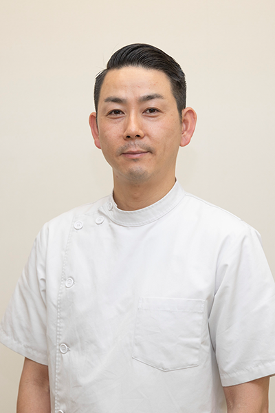
もし、お近くにお住まいで、困っているならば、一度ひまわり接骨院までお問い合わせください。腰痛・坐骨神経痛の専門家の新幡が、ご相談に乗ります。
気軽にご相談ください。

新着情報・お知らせ
平常通り営業致します。
日・祝休みです。
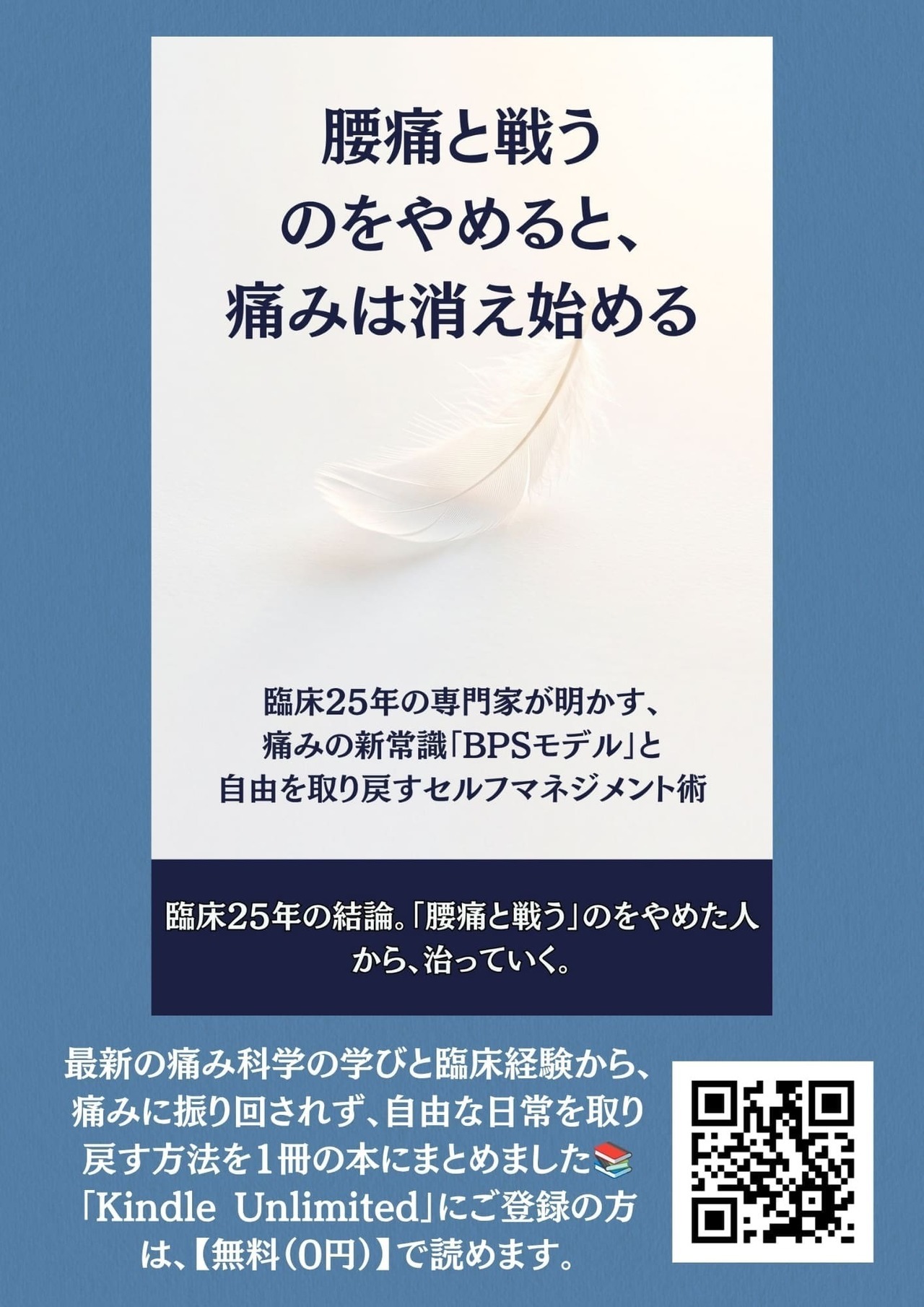
タイトル「腰痛は戦うのをやめると、痛みは消え始める」
Amazonの「Kindle Unlimited」にご登録の方は、【無料(0円)】で今すぐお読みいただけます!
↓↓
こちらから
タイトル「不安から逃げるのをやめると、腰痛は消え始める」」
Amazonの「Kindle Unlimited」にご登録の方は、【無料(0円)】で今すぐお読みいただけます!
↓↓
こちらから

お気軽にお問合せください

お電話でのお問合せ・相談予約
<受付時間>
月~土
9:00〜13:00 /15:00〜19:00
※日曜・祝日は除く
フォームは24時間受付中です。お気軽にご連絡ください。
ひまわり接骨院
住所
〒213-0002
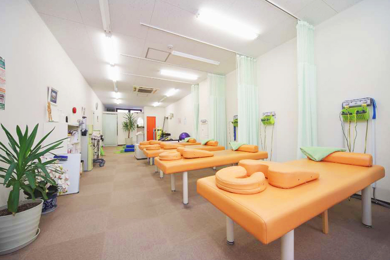
神奈川県川崎市高津区二子1丁目7−17 リバーサイドマンション杉崎 102
アクセス
二子新地駅 徒歩3分
駐車場:近隣にコインパーキングあり。自転車・バイクは店舗前に駐輪場がございます。
受付時間
月~土
9:00〜13:00 /15:00〜19:00
定休日
日曜・祝日